. 2022 Mar 2:2022:5739909.
doi: 10.1155/2022/5739909. eCollection 2022.

Fuzheng Jiedu Decoction Induces Apoptosis and Enhances Cisplatin Efficacy in Ovarian Cancer Cells In Vitro and In Vivo through Inhibiting the PI3K/AKT/mTOR/NF- κ B Signaling Pathway

Affiliations

Fuzheng Jiedu Decoction Induces Apoptosis and Enhances Cisplatin Efficacy in Ovarian Cancer Cells In Vitro and In Vivo through Inhibiting the PI3K/AKT/mTOR/NF- κ B Signaling Pathway

Huadi Yang et al. Biomed Res Int. .

Abstract

Objectives: This study is aimed at investigating the anticancer activity of Fuzheng Jiedu decoction (FJD) alone or in combination with cisplatin in ovarian cancer (OC) models, as well as its underlying mechanisms of action.

Methods: The anticancer activities of FJD, cisplatin, and the combination of the PI3K inhibitor (LY294002, LY) or activator (IGF-1) were evaluated in OC cell lines in vitro and in a SKOV3 xenograft mouse model in vivo. The cell proliferation and invasion ability were measured using MTT, EdU, and transwell assays, respectively. The cell apoptosis was examined by flow cytometry and JC-1 assays. The expression levels of the Bcl-2 family and the PI3K/AKT/mTOR/NF-κB pathway-related proteins were analyzed by Western blot.

Results: The in vivo and in vitro studies showed that FJD administration could significantly inhibit cell proliferation and promote cell apoptosis in two OC cell lines SKOV3 and 3AO and partially decreased the tumor volumes and weights. In addition, FJD could significantly downregulate the protein levels of p-PI3K/PI3K, p-AKT/AKT, p-mTOR/mTOR, NF-κB, p38, and Bcl-2 and upregulate the Bax, Cyt-C, and cleaved caspase-3 in OC tumor tissues and cells. FJD cotreatment increased the efficacy of cisplatin, including inhibiting OC cell proliferation and invasion, promoting cell apoptosis, and inhibiting the PI3K/AKT/mTOR signaling pathway, while this enhancement was suppressed by IGF-1. Similarly, LY also enhanced the anticancer efficacy of cisplatin.

Conclusions: This study indicated that FJD could improve the efficacy of cisplatin by inhibiting the PI3K/AKT/mTOR/NF-κB signaling pathway. It is suggested that FJD may be a valuable adjuvant drug for the treatment of OC.


Conflict of interest statement

The authors declare that they have no conflicts of interest.

Figures

Figure 1
Figure 1
Chemical components of Fuzheng Jiedu decoction (FJD): (a) UPLC-Q/TOF-MS analysis in a positive mode; (b) UPLC-Q/TOF-MS analysis in a negative mode.
Figure 2
Figure 2
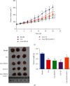
Effects of different doses of FJD on tumor volumes and weights and histopathology in OC tumor-bearing mice: (a) tumor volumes; (b) tumor weights; (c) HE staining of tumor tissues. Data was expressed as mean ± SD.
Figure 3
Figure 3
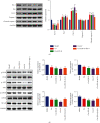
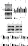
Effects of different doses of FJD on the Bcl-2 family protein (a) and the PI3K/AKT/mTOR/NF-κB (b) signaling pathway of protein levels in tumor tissues. Data was expressed as mean ± SD. Compared with the model group, P < 0.05, ★★P < 0.01.
Figure 4
Figure 4
Cisplatin inhibited tumor volumes and weights and FJD enhancement effect in mice: (a) tumor volumes and (b) tumor weights. Data was expressed as mean ± SD. Compared with the model group, P < 0.05, ★★P < 0.01; compared with the Cis group, #P < 0.05.
Figure 5
Figure 5
Regulatory effect of FJD combined with cisplatin on apoptosis-related proteins (a) and the PI3K/AKT/mTOR/NF-κB (b) signaling pathway in the animal model. Data was expressed as mean ± SD. Compared with the model group, P < 0.05, ★★P < 0.01; compared with the Cis+FJD-H group, %P < 0.05.
Figure 6
Figure 6
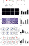
FJD inhibited the cell proliferation, invasion, and apoptosis of two OC cell lines SKOV-3 and 3AO: (a) time variation in viability of SKOV-3 and 3AO cells; (b) EdU assessed cell proliferation toxicity; (c) changes of invasion ability of SKOV3 and 3AO cells in each group (200x); (d) the cell apoptosis was detected by the Annexin V-FITC/PI staining with flow cytometry; (e) the mitochondrial transmembrane potential was detected by the JC-1 staining with flow cytometry. Data was expressed as mean ± SD. Compared with the control group, P < 0.05, ▲▲P < 0.01; compared with the FJD-H group, P < 0.05, ∗∗P < 0.01).
Figure 7
Figure 7
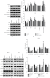
FJD p6romoted OC cell apoptosis by regulating the PI3K/AKT/mTOR/P38 signaling pathway: (a, b) the expressions of Bax, Cyt-C, caspase-3, cleaved caspase-3, and Bcl-2 in OC cells; (c, d) the protein levels of p-PI3K/PI3K, p-AKT/AKT, p-mTOR/mTOR, and p38 in SKOV3 and 3AO cells, respectively. Data was expressed as mean ± SD. Compared with the control group, P < 0.05, ▲▲P < 0.01; compared with the FJD-H group, P < 0.05, ∗∗P < 0.01.
Figure 8
Figure 8
Inhibitory effect of cisplatin on the cell growth, invasion, and apoptosis of SKOV-3 and 3AO cells and FJD enhancing the efficacy of cisplatin: (a) the cell viability of SKOV-3 and 3AO cells; (b) EdU assessed cell proliferation toxicity; (c) the changes of cell invasion ability; (d) the cell apoptosis was detected by the Annexin V-FITC/PI staining with flow cytometry; (e) the mitochondrial transmembrane potential was detected by the JC-1 staining with flow cytometry. Data was expressed as mean ± SD. Compared with the control group, P < 0.05, ▲▲P < 0.01; compared with the Cis group, #P < 0.05, ##P < 0.05; compared with the Cis+FJD-H group, %P < 0.05, %%P < 0.01.
Figure 9
Figure 9
Effect of FJD combined with cisplatin on the apoptosis and the PI3K/AKT/mTOR signaling pathway at the cell level: (a, b) the expressions of Bax, Cyt-C, caspase-3, cleaved caspase-3, and Bcl-2 in two OC cell lines; (c, d) the protein levels of p-PI3K/PI3K, p-AKT/AKT, and p-mTOR/mTOR in SKOV3 and 3AO cells were detected, respectively. Data was expressed as mean ± SD. Compared with the control group, P < 0.05, ▲▲P < 0.01; compared with the Cis group, #P < 0.05, ##P < 0.05; compared with the Cis+FJD-H group, %P < 0.05.

Similar articles

Cited by

References

    1. Rossi L., Verrico M., Zaccarelli E., et al. Bevacizumab in ovarian cancer: a critical review of phase III studies. Oncotarget . 2017;8(7):12389–12405. doi: 10.18632/oncotarget.13310. - DOI - PMC - PubMed
    1. Erratum. Erratum: Global cancer statistics 2018: GLOBOCAN estimates of incidence and mortality worldwide for 36 cancers in 185 countries. CA: a Cancer Journal for Clinicians . 2020;70(4):p. 313. doi: 10.3322/caac.21609. - DOI - PubMed
    1. De Angelis R., Sant M., Coleman M. P., et al. Cancer survival in Europe 1999-2007 by country and age: results of EUROCARE--5-a population-based study. The Lancet. Oncology . 2014;15(1):23–34. doi: 10.1016/S1470-2045(13)70546-1. - DOI - PubMed
    1. Yang J., Nie J., Ma X., Wei Y., Peng Y., Wei X. Targeting PI3K in cancer: mechanisms and advances in clinical trials. Molecular Cancer . 2019;18(1):p. 26. doi: 10.1186/s12943-019-0954-x. - DOI - PMC - PubMed
    1. Ediriweera M. K., Tennekoon K. H., Samarakoon S. R. Role of the PI3K/AKT/mTOR signaling pathway in ovarian cancer: biological and therapeutic significance. Seminars in Cancer Biology . 2019;59:147–160. doi: 10.1016/j.semcancer.2019.05.012. - DOI - PubMed

MeSH terms