

Molecular Mechanisms of Immunosenescene and Inflammaging: Relevance to the Immunopathogenesis and Treatment of Multiple Sclerosis
- PMID: 35281989
- PMCID: PMC8913495
- DOI: 10.3389/fneur.2021.811518
Molecular Mechanisms of Immunosenescene and Inflammaging: Relevance to the Immunopathogenesis and Treatment of Multiple Sclerosis
Abstract
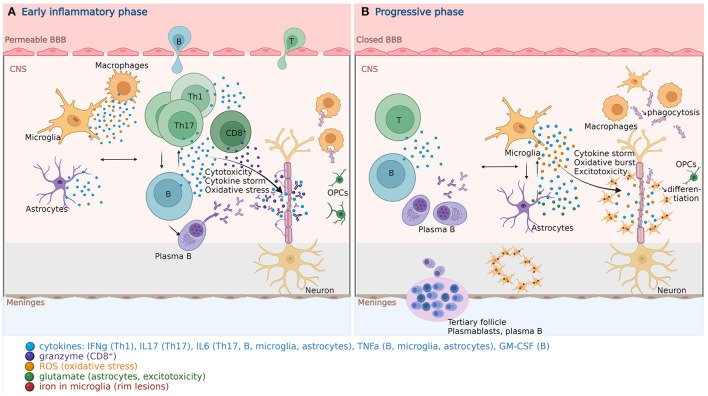
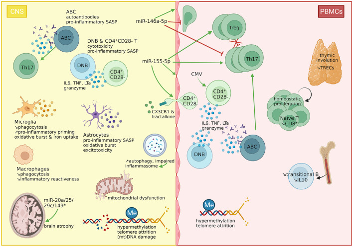
Aging is characterized, amongst other features, by a complex process of cellular senescence involving both innate and adaptive immunity, called immunosenescence and associated to inflammaging, a low-grade chronic inflammation. Both processes fuel each other and partially explain increasing incidence of cancers, infections, age-related autoimmunity, and vascular disease as well as a reduced response to vaccination. Multiple sclerosis (MS) is a lifelong disease, for which considerable progress in disease-modifying therapies (DMTs) and management has improved long-term survival. However, disability progression, increasing with age and disease duration, remains. Neurologists are now involved in caring for elderly MS patients, with increasing comorbidities. Aging of the immune system therefore has relevant implications for MS pathogenesis, response to DMTs and the risks mediated by these treatments. We propose to review current evidence regarding markers and molecular mechanisms of immunosenescence and their relevance to understanding MS pathogenesis. We will focus on age-related changes in the innate and adaptive immune system in MS and other auto-immune diseases, such as systemic lupus erythematosus and rheumatoid arthritis. The consequences of these immune changes on MS pathology, in interaction with the intrinsic aging process of central nervous system resident cells will be discussed. Finally, the impact of immunosenescence on disease evolution and on the safety and efficacy of current DMTs will be presented.
Keywords: T/B cells; astrocytes; disease modifying therapies; immunosenescence; inflammaging; microglia; multiple sclerosis; oligodendrocytes.
Copyright © 2022 Perdaens and van Pesch.
Conflict of interest statement
The authors declare that the research was conducted in the absence of any commercial or financial relationships that could be construed as a potential conflict of interest.
Figures
Similar articles
-
Immunosenescence and multiple sclerosis: inflammaging for prognosis and therapeutic consideration.Front Aging. 2023 Oct 13;4:1234572. doi: 10.3389/fragi.2023.1234572. eCollection 2023. Front Aging. 2023. PMID: 37900152 Free PMC article. Review.
-
Immunosenescence and multiple sclerosis.Neurol Neurochir Pol. 2022;56(3):220-227. doi: 10.5603/PJNNS.a2022.0045. Epub 2022 Jun 23. Neurol Neurochir Pol. 2022. PMID: 35735245
-
The Impact of Aging on Multiple Sclerosis.Curr Neurol Neurosci Rep. 2024 Apr;24(4):83-93. doi: 10.1007/s11910-024-01333-2. Epub 2024 Feb 28. Curr Neurol Neurosci Rep. 2024. PMID: 38416310 Review.
-
The Role of Immunosenescence in Cerebral Small Vessel Disease: A Review.Int J Mol Sci. 2022 Jun 27;23(13):7136. doi: 10.3390/ijms23137136. Int J Mol Sci. 2022. PMID: 35806140 Free PMC article. Review.
-
Contributions of Age-Related Thymic Involution to Immunosenescence and Inflammaging.Immun Ageing. 2020 Jan 20;17:2. doi: 10.1186/s12979-020-0173-8. eCollection 2020. Immun Ageing. 2020. PMID: 31988649 Free PMC article. Review.
Cited by
-
From aging to long COVID: exploring the convergence of immunosenescence, inflammaging, and autoimmunity.Front Immunol. 2023 Oct 24;14:1298004. doi: 10.3389/fimmu.2023.1298004. eCollection 2023. Front Immunol. 2023. PMID: 37942323 Free PMC article. Review.
-
Selective Upregulation of SIRT1 Expression in Retinal Ganglion Cells by AAV-Mediated Gene Delivery Increases Neuronal Cell Survival and Alleviates Axon Demyelination Associated with Optic Neuritis.Biomolecules. 2022 Jun 14;12(6):830. doi: 10.3390/biom12060830. Biomolecules. 2022. PMID: 35740955 Free PMC article.
-
Microglia activation in periplaque white matter in multiple sclerosis depends on age and lesion type, but does not correlate with oligodendroglial loss.Acta Neuropathol. 2023 Dec;146(6):817-828. doi: 10.1007/s00401-023-02645-2. Epub 2023 Oct 28. Acta Neuropathol. 2023. PMID: 37897549 Free PMC article.
-
Aging in multiple sclerosis: from childhood to old age, etiopathogenesis, and unmet needs: a narrative review.Front Neurol. 2023 Jun 2;14:1207617. doi: 10.3389/fneur.2023.1207617. eCollection 2023. Front Neurol. 2023. PMID: 37332984 Free PMC article. Review.
-
The Protective Effect of Astragalus Polysaccharide on Experimental Autoimmune Encephalomyelitis in Mice by Activating the AMPK/JAK/ STAT3/Arginase-1 Signaling Pathway.Curr Pharm Biotechnol. 2025;26(6):863-871. doi: 10.2174/0113892010314302240902073112. Curr Pharm Biotechnol. 2025. PMID: 39289935
References
-
- United Nations Department Department of Economic and Social Affairs Population Division . World Population Prospects 2019, Volume II: Demographic Profiles. New York, NY: (2019).
Publication types
LinkOut - more resources
Full Text Sources