. 2022 Feb 23:12:740603.
doi: 10.3389/fneur.2021.740603. eCollection 2021.

Automated Quantification of Brain Lesion Volume From Post-trauma MR Diffusion-Weighted Images

Affiliations

Automated Quantification of Brain Lesion Volume From Post-trauma MR Diffusion-Weighted Images

Thomas Mistral et al. Front Neurol. .

Abstract

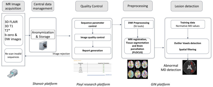
Objectives: Determining the volume of brain lesions after trauma is challenging. Manual delineation is observer-dependent and time-consuming and cannot therefore be used in routine practice. The study aimed to evaluate the feasibility of an automated atlas-based quantification procedure (AQP) based on the detection of abnormal mean diffusivity (MD) values computed from diffusion-weighted MR images.

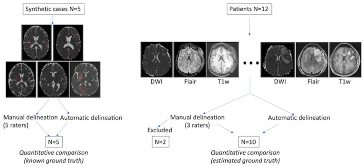
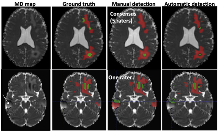
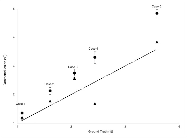
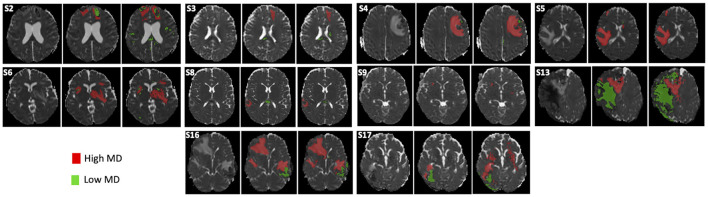
Methods: The performance of AQP was measured against manual delineation consensus by independent raters in two series of experiments based on: (i) realistic trauma phantoms (n = 5) where low and high MD values were assigned to healthy brain images according to the intensity, form and location of lesion observed in real TBI cases; (ii) severe TBI patients (n = 12 patients) who underwent MR imaging within 10 days after injury.

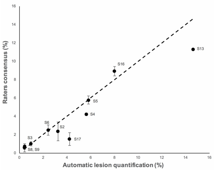
Results: In realistic TBI phantoms, no statistical differences in Dice similarity coefficient, precision and brain lesion volumes were found between AQP, the rater consensus and the ground truth lesion delineations. Similar findings were obtained when comparing AQP and manual annotations for TBI patients. The intra-class correlation coefficient between AQP and manual delineation was 0.70 in realistic phantoms and 0.92 in TBI patients. The volume of brain lesions detected in TBI patients was 59 ml (19-84 ml) (median; 25-75th centiles).

Conclusions: Our results support the feasibility of using an automated quantification procedure to determine, with similar accuracy to manual delineation, the volume of low and high MD brain lesions after trauma, and thus allow the determination of the type and volume of edematous brain lesions. This approach had comparable performance with manual delineation by a panel of experts. It will be tested in a large cohort of patients enrolled in the multicenter OxyTC trial (NCT02754063).

Keywords: MRI; brain; mean diffusion (MD); segmentation (image processing); traumatic brain injury.


Conflict of interest statement

The authors declare that the research was conducted in the absence of any commercial or financial relationships that could be construed as a potential conflict of interest.

Figures

Figure 1
Figure 1
Evaluation procedure. Left: five realistic TBI lesion cases were constructed with low (green) and high (red) artificial MD values. The ground truth was predefined for automated and manual lesion delineation comparison. Right: Twelve TBI patients were included, each with three types of MR image. Manual and automated delineation results were quantitatively compared for 10 patients. The ground truth was defined as the consensus of expert annotations (“consensual inter-raters ground truth”), calculated using STAPLE (10).
Figure 2
Figure 2
Image processing pipeline from image acquisition to automated detection of mean diffusivity abnormalities.
Figure 3
Figure 3
Typical examples of abnormal mean diffusivity (MD) values introduced in diffusion-weighted images (DWI) of two healthy volunteers (realistic TBI phantoms). Top: Good agreement between manual and automated segmentation. Bottom: Moderate agreement between manual and automated segmentation. The artifact (white arrow) was falsely detected as a lesion by one rater. Red, High MD values; Green, Low MD values.
Figure 4
Figure 4
The correspondence analysis in brain lesion volume for the five realistic phantom cases for both the raters' consensus (circle) and AQP method (triangle) (y-axis) vs. the ground truth (x-axis). Total lesion volume (low + high MD) in % brain volume of diffusion-weighted images (mean, 95% confidence interval). The dashed line indicates the identity curve.
Figure 5
Figure 5
Delineation of brain lesions from diffusion-weighted images (DWI) in 10 TBI patients. The MD map (left), rater consensus (middle) and automated quantification procedure (right) is shown for each patient. S2–S17 refer to the corresponding TBI subject (see Table 3).
Figure 6
Figure 6
The correspondence analysis in brain lesion volume for the then TBI cases for both the raters' consensus (y-axis) and AQP method (y-axis). Total lesion volume (low + high MD) in % of the brain volume of diffusion-weighted images (mean, 95% confidence interval). The dashed line indicates the identity curve.

References

    1. Thornhill S, Teasdale GM, Murray GD, McEwen J, Roy CW, Penny KI. Disability in young people and adults one year after head injury: prospective cohort study. BMJ. (2000) 320:1631–5. 10.1136/bmj.320.7250.1631 - DOI - PMC - PubMed
    1. Shenton ME, Hamoda HM, Schneiderman JS, Bouix S, Pasternak O, Rathi Y, et al. . A review of magnetic resonance imaging and diffusion tensor imaging findings in mild traumatic brain injury. Brain Imaging Behav. (2012) 6:137–92. 10.1007/s11682-012-9156-5 - DOI - PMC - PubMed
    1. Pasco A, Ter Minassian A, Chapon C, Lemaire L, Franconi F, Darabi D, et al. . Dynamics of cerebral edema and the apparent diffusion coefficient of water changes in patients with severe traumatic brain injury. A prospective MRI study. Eur Radiol. (2006) 16:1501–8. 10.1007/s00330-005-0086-0 - DOI - PubMed
    1. Galanaud D, Perlbarg V, Gupta R, Stevens RD, Sanchez P, Tollard E, et al. . Assessment of white matter injury and outcome in severe brain trauma a prospective multicenter cohort. Anesthesiology. (2012) 117:1300–10. 10.1097/ALN.0b013e3182755558 - DOI - PubMed
    1. Patel A, van Ginneken B, Meijer FJA, van Dijk EJ, Prokop M, Manniesing R. Robust cranial cavity segmentation in CT and CT perfusion images of trauma and suspected stroke patients. Med Image Anal. (2017) 36:216–28. 10.1016/j.media.2016.12.002 - DOI - PubMed

Associated data