

A novel de novo TBX20 variant in a 6-year-old Chinese girl with left ventricular noncompaction: a case report
- PMID: 35282022
- PMCID: PMC8905101
- DOI: 10.21037/tp-21-460
A novel de novo TBX20 variant in a 6-year-old Chinese girl with left ventricular noncompaction: a case report
Abstract
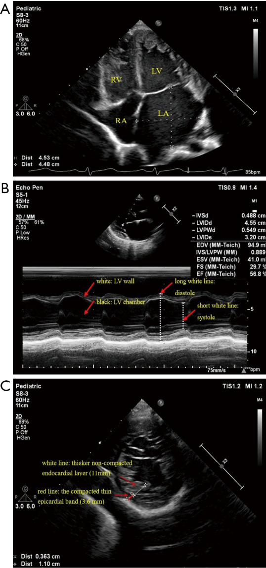
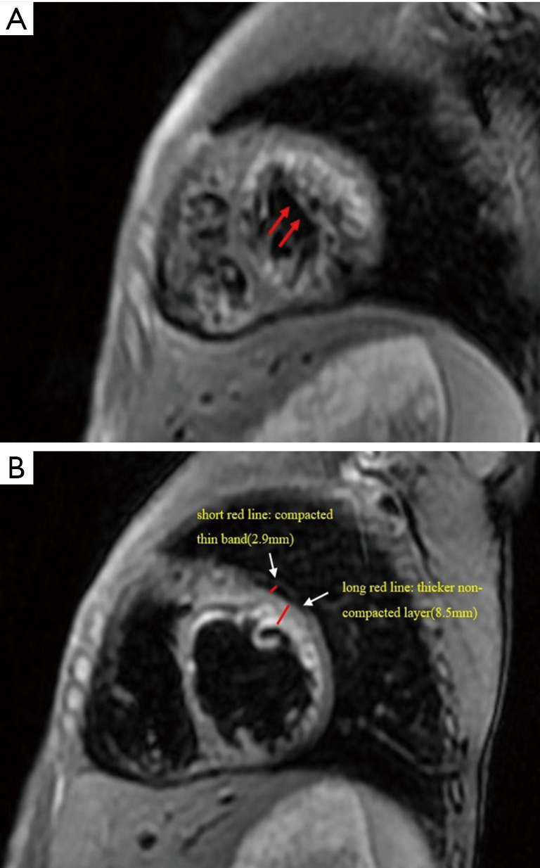
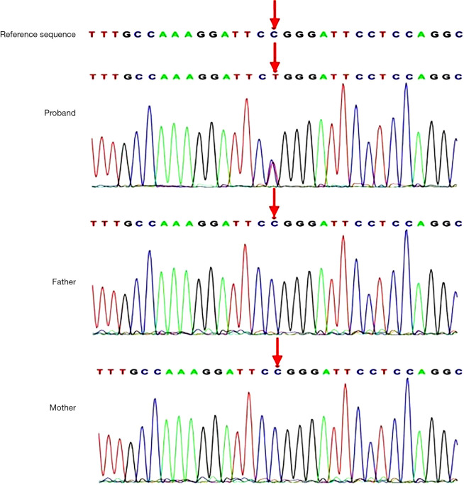
Left ventricular noncompaction (LVNC) is a particular type of cardiomyopathy with an excessively prominent trabecular meshwork and deep intertrabecular recesses in the left ventricle (LV). The clinical manifestation of LVNC is highly variable, ranging from no symptom to congestive heart failure, arrhythmia, thrombosis, and potentially sudden cardiac death. Approximately half of LVNC cases are hereditary. TBX20 is expressed in human embryonic and vertebrate hearts. In this article, we report on a case of pediatric LVNC with a novel de novo TBX20 [c.859C>T, p.(Arg287Trp)] gene variant, which appears to be pathogenic and had not been previously reported in LVNC. The 6-year-old girl was admitted to our hospital for unexplained syncope. 2D-echocardiography revealed a dilated LV with numerous prominent trabeculations, and a two-layered structure, comprising a compacted thin epicardial band and a thicker non-compacted endocardial layer, with deep endomyocardial spaces and intertrabecular recesses in LV. During the follow-up, the child has not shown any obvious clinical signs or symptoms. In this case report, the de novo variant of TBX20 in LVNC expands the spectrum of variants that cause LVNC and contributes to the genetic counseling and individualized treatment of patients. Clinicians should focus on exploring the clinical and genetic characteristics of LVNC to provide therapies and follow-up to improve the outcome.
Keywords: TBX20; case report; exome sequencing (ES); left ventricular noncompaction (LVNC).
2022 Translational Pediatrics. All rights reserved.
Conflict of interest statement
Conflicts of Interest: All authors have completed the ICMJE uniform disclosure form (available at https://tp.amegroups.com/article/view/10.21037/tp-21-460/coif). The authors have no conflicts of interest to declare.
Figures
Similar articles
-
Dilated-Left Ventricular Non-Compaction Cardiomyopathy in a Pediatric Case with SPEG Compound Heterozygous Variants.Int J Mol Sci. 2022 May 6;23(9):5205. doi: 10.3390/ijms23095205. Int J Mol Sci. 2022. PMID: 35563595 Free PMC article.
-
Left ventricular noncompaction - Risk stratification and genetic consideration.J Cardiol. 2020 Jan;75(1):1-9. doi: 10.1016/j.jjcc.2019.09.011. Epub 2019 Oct 17. J Cardiol. 2020. PMID: 31629663 Review.
-
The TNNI3 Arg192His mutation in a 13-year-old girl with left ventricular noncompaction.J Cardiol Cases. 2018 Jul 1;18(1):33-36. doi: 10.1016/j.jccase.2018.04.001. eCollection 2018 Jul. J Cardiol Cases. 2018. PMID: 30279906 Free PMC article.
-
Role of TBX20 Truncating Variants in Dilated Cardiomyopathy and Left Ventricular Noncompaction.Circ Genom Precis Med. 2024 Apr;17(2):e004404. doi: 10.1161/CIRCGEN.123.004404. Epub 2024 Feb 14. Circ Genom Precis Med. 2024. PMID: 38353104 Free PMC article.
-
Myocardial Mechanics and Associated Valvular and Vascular Abnormalities in Left Ventricular Noncompaction Cardiomyopathy.J Clin Med. 2023 Dec 22;13(1):78. doi: 10.3390/jcm13010078. J Clin Med. 2023. PMID: 38202085 Free PMC article. Review.
References
-
- Maron BJ, Towbin JA, Thiene G, et al. Contemporary definitions and classification of the cardiomyopathies: an American Heart Association scientific statement from the council on clinical cardiology, heart failure and transplantation committee; quality of care and outcomes research and functional genomics and translational biology interdisciplinary working groups; and council on epidemiology and prevention. Circulation 2006;113:1807-16. 10.1161/CIRCULATIONAHA.106.174287 - DOI - PubMed
-
- Richards S, Aziz N, Bale S, et al. Standards and guidelines for the interpretation of sequence variants: a joint consensus recommendation of the American College of Medical Genetics and Genomics and the Association for Molecular Pathology. Genet Med 2015;17:405-24. 10.1038/gim.2015.30 - DOI - PMC - PubMed
Publication types
LinkOut - more resources
Full Text Sources