Review
. 2022 Feb 24:9:836473.
doi: 10.3389/fcvm.2022.836473. eCollection 2022.

Non-invasive Multimodality Imaging of Coronary Vulnerable Patient

Affiliations
Review

Non-invasive Multimodality Imaging of Coronary Vulnerable Patient

Marjorie Canu et al. Front Cardiovasc Med. .

Abstract

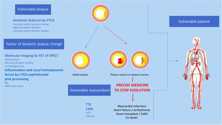
Atherosclerotic plaque rupture or erosion remain the primary mechanism responsible for myocardial infarction and the major challenge of cardiovascular researchers is to develop non-invasive methods of accurate risk prediction to identify vulnerable plaques before the event occurs. Multimodal imaging, by CT-TEP or CT-SPECT, provides both morphological and activity information about the plaque and cumulates the advantages of anatomic and molecular imaging to identify vulnerability features among coronary plaques. However, the rate of acute coronary syndromes remains low and the mechanisms leading to adverse events are clearly more complex than initially assumed. Indeed, recent studies suggest that the detection of a state of vulnerability in a patient is more important than the detection of individual sites of vulnerability as a target of focal treatment. Despite this evolution of concepts, multimodal imaging offers a strong potential to assess patient's vulnerability. Here we review the current state of multimodal imaging to identify vulnerable patients, and then focus on emerging imaging techniques and precision medicine.

Keywords: chronic coronary syndrome; coronary artery disease; multimodal imaging; risk stratification; vulnerable patient; vulnerable plaque.


Conflict of interest statement

The authors declare that the research was conducted in the absence of any commercial or financial relationships that could be construed as a potential conflict of interest.

Figures

Figure 1
Figure 1
Example of a patient reporting exertional dyspnea. In (A), CTCA showed significant CAD on left anterior descending (LAD) artery (white arrow), classified CAD-RADS 4A. Coronary angiogram confirm severe stenosis of proximal LAD (black arrow), angioplasty followed by stenting was performed to relieve symptoms (B). Finally, CTCA post-treatment based on the FAI-Score values (C) on three arteries, the coronary atherosclerotic plaque burden and the clinical risk factors showed low CaRi-Heart Risk, thereby predicting low risk of future acute coronary events and permitted treatment goals and follow-up strategies personalization.
Figure 2
Figure 2
Proposition of non-invasive multimodality imaging strategy to detect and treat coronary vulnerable patient. CTCA, Computed tomography coronary angiography; CV, cardiovascular; CMR, cardiac magnetic resonance imaging, FAI, fat attenuation index; LVEF, left ventricular ejection fraction; PET, positron emission tomography; SPECT, single photon emission computed tomography; TTE, trans thoracic echocardiography.

References

    1. Boden WE, O'Rourke RA, Teo KK, Hartigan PM, Maron DJ, Kostuk WJ, et al. . Optimal medical therapy with or without PCI for stable coronary disease. N Engl J Med. (2007) 356:1503–16. 10.1056/NEJMoa070829 - DOI - PubMed
    1. Rutter MK, Nesto RW. The BARI 2D study: a randomised trial of therapies for type 2 diabetes and coronary artery disease. Diab Vasc Dis Res. (2010) 7:69–72. 10.1177/1479164109354145 - DOI - PubMed
    1. Libby P, Theroux P. Pathophysiology of coronary artery disease. Circulation. (2005) 111:3481–8. 10.1161/CIRCULATIONAHA.105.537878 - DOI - PubMed
    1. Stone GW, Maehara A, Lansky AJ, de Bruyne B, Cristea E, Mintz GS, et al. . A prospective natural-history study of coronary atherosclerosis. N Engl J Med. (2011) 364:226–35. 10.1056/NEJMoa1002358 - DOI - PubMed
    1. Virmani R, Kolodgie FD, Burke AP, Farb A, Schwartz SM. Lessons from sudden coronary death: a comprehensive morphological classification scheme for atherosclerotic lesions. Arterioscler Thromb Vasc Biol. (2000) 20:1262–7. 10.1161/01.ATV.20.5.1262 - DOI - PubMed

LinkOut - more resources