. 2022 Mar 3:26:101411.
doi: 10.1016/j.ajoc.2022.101411. eCollection 2022 Jun.

Multimodal retinal imaging of m.3243A>G associated retinopathy

Affiliations

Multimodal retinal imaging of m.3243A>G associated retinopathy

Francesco Romano et al. Am J Ophthalmol Case Rep. .

Abstract

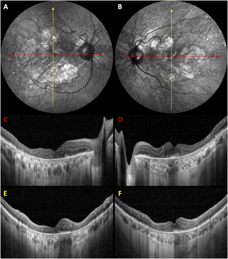
Retro-mode illumination imaging can provide good visualization of chorio-retinal atrophy and of the retinal pigment epithelial alterations occurring in m.3243A > G associated retinopathy.

Keywords: Chorio-retinal atrophy; Encephalopathy; Lactic acidosis and stroke-like episodes syndrome; MELAS; MELAS, mitochondrial myopathy, encephalopathy, lactic acidosis and stroke-like episodes; MTTL1 gene; Mitochondrial myopathy; Mitochondrial retinopathy; Multimodal imaging; NIR-AF, near-infrared autofluorescence; OCT, optical coherence tomography; RPE, retinal pigment epithelium; Retro-mode illumination; m.3243A>G.


Conflict of interest statement

MC has the following disclosures: Bayer (recipient), Nidek (recipient). FR and APS have no financial disclosures. GS is consultant for Heidelberg Engineering, Optos, OptoVue, CenterVue, Allergan, Bayer, Genetech, Novartis, Quantel Medical, Carl Zeiss Meditec, Boheringer, Topcon, Roche.

Figures

Fig. 1
Fig. 1
Multimodal retinal imaging of m.3243A > G associated retinopathy. A– B: Color fundus photographs (Eidon; CenterVue, Padua, Italy) show large areas of chorio-retinal atrophy with minimal foveal sparing, bordered by numerous yellowish sub-retinal lesions that extend to the nasal peripapillary area. C– D: The widefield fundus autofluorescence images (55° × 55°, Spectralis HRA2; Heidelberg Engineering GmbH, Heidelberg, Germany) confirms the presence of retinal pigment epithelium (RPE) atrophy surrounded by a granular pattern of hypo- and hyper-autofluorescence.E– F: Near-infrared autofluorescence signal appears similarly decreased in the atrophic areas and the surrounding RPE alterations with a better visualization of the underlying hypo-autofluorescent choroidal vessels; interestingly, the foveal hyper-autofluorescent spot results partially preserved in both eyes. G– H: Retro-mode illumination imaging (Mirante; Nidek co. Ltd, Gamagori, Japan) further refines the RPE irregularities around the macular atrophy, corresponding to the sub-retinal deposits.
Fig. 2
Fig. 2
Near-infrared reflectance images (A–B) and optical coherence tomography (OCT, C–F). Structural OCT shows extensive and well-demarcated retinal pigment epithelium atrophy with numerous outer retinal tubulations lying above the Bruch's membrane in horizontal scans (C,D). The foveal ellipsoid and interdigitation zones appear significantly thickened in the preserved areas (D,E). Sub-retinal flecks/spots can be better appreciated in vertical OCT scans (E,F).

References

    1. Coussa R.G., Parikh S., Traboulsi E.I. Mitochondrial DNA A3243G variant-associated retinopathy: current perspectives and clinical implications. Surv Ophthalmol. 2021;66(5):838–855. doi: 10.1016/j.survophthal.2021.02.008.PMID:33610586. - DOI - PubMed
    1. de Laat P., Smeitink J.A.M., Janssen M.C.H., et al. Mitochondrial retinal dystrophy associated with the m.3243A>G mutation. Ophthalmology. 2013;120(12):2684–2696. doi: 10.1016/j.ophtha.2013.05.013.PMID:23806424. - DOI - PubMed
    1. Birtel J., von Landenberg C., Gliem M., et al. Mitochondrial retinopathy. Ophthalmol Retina. 2021 doi: 10.1016/j.oret.2021.02.017. S2468-6530(21)00071-3, PMID: 34257060. - DOI - PubMed