. 2022 Jan 20;13(6):1780-1788.
doi: 10.1039/d1sc05204g. eCollection 2022 Feb 9.

Site-specific DNA functionalization through the tetrazene-forming reaction in ionic liquids

Affiliations

Site-specific DNA functionalization through the tetrazene-forming reaction in ionic liquids

Seiya Ishizawa et al. Chem Sci. .

Erratum in

Abstract

Development of multiple chemical tools for deoxyribonucleic acid (DNA) labeling has facilitated wide use of their functionalized conjugates, but significant practical and methodological challenges remain to achievement of site-specific chemical modification of the biomacromolecule. As covalent labeling processes are more challenging in aqueous solution, use of nonaqueous, biomolecule-compatible solvents such as an ionic liquid consisting of a salt with organic molecule architecture, could be remarkably helpful in this connection. Herein, we demonstrate site-specific chemical modification of unprotected DNAs through a tetrazene-forming amine-azide coupling reaction using an ionic liquid. This ionic liquid-enhanced reaction process has good functional group tolerance and precise chemoselectivity, and enables incorporation of various useful functionalities such as biotin, cholesterol, and fluorophores. A site-specifically labeled oligonucleotide, or aptamer interacting with a growth factor receptor (Her2) was successfully used in the fluorescence imaging of breast cancer cell lines. The non-traditional medium-promoted labeling strategy described here provides an alternative design paradigm for future development of chemical tools for applications involving DNA functionalization.


Conflict of interest statement

The authors declare the following competing financial interest(s): the authors have filed a patent application on the ionic-liquid-based tetrazene-forming bioconjugation method.

Figures

Fig. 1
Fig. 1. Site-specific tetrazene-forming reaction on DNA substrates.
Fig. 2
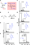
Fig. 2. Alkylamine-selective tetrazene-forming reaction with pentanucleotides in an ionic liquid. Reaction conditions: KHCO3 (20 mM), XTTTT (0.2 mM), azide 1a (7.5 mM), and PPh3 (20 mM) in 1-butyl-1-methylpyrrolidinium trifluoromethanesulfonate (BMPy OTf) at 50 °C for 2 h. (A) Chemical structure of nucleotide backbone in an XTTTT sequence with or without an alkylamine. (B) Chemical structure of azide 1a. (C) Structure of the ionic liquid, BMPy OTf. (D–I) Matrix-assisted laser desorption/ionization (MALDI-MS) analysis of the reaction of XTTTT where X = adenosine (D), thymidine (E), cytidine (F), guanosine (G), deoxyuridine (H), and thymidine with alkylamine (I) containing a 12 carbon linker (5AmMC12).
Fig. 3
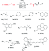
Fig. 3. Tetrazene-forming DNA bioconjugation with a variety of alkylazides. Modification reaction conditions: KHCO3 (20 mM), 5′-TTTTT-3′-alkyl-NH2 (0.2 mM), azide derivatives 1b–1j (7.5 mM), and PPh3 (20 mM) in BMPy OTf at 50 °C for 2 h. *Reaction was incubated overnight. Conversion in the parentheses were calculated based on matrix-assisted laser desorption/ionization (MALDI-MS) analysis. Conversion obtained by liquid chromatography is available in Fig. S2.
Fig. 4
Fig. 4. Screening of phosphine reagents on tetrazene formation reaction on a DNA aptamer toward human serum albumin (HSA). Sequence of the HSA aptamer: 5′-TGCGGTTGTAGTACTCGTGGCCG-3′. Reaction conditions: HSA DNA aptamer (0.1 mM), KHCO3 (20 mM), biotin azide (7.5 mM), and phosphine (3.0 mM) in a mixture of 1-ethyl-3-methylimidazolium acetate (EMIM OAc)/DMF (1 : 1) at 50 °C for 2 h. (A) General chemical structure of phosphine. Green circle, red square, and blue triangle each represents the aryl or alkyl groups shown in Fig. 5C. (B) Chemical structure of ionic liquid EMIM OAc. (C) Chemical structures of the aryl or alkyl groups of different phosphine/phosphite reagents. (D) Bar graph showing the anti-biotin Southern blot after modification of the HSA DNA aptamer with biotin azide with different phosphines. Error bars represent standard deviation (n = 3). Representative blot membrane images for the anti-biotin Southern blot (Cy5) and total stain with Mayer's hemalum solution (MHS) are shown below the bar graphs. Full-width Southern blot membrane images are shown in Fig. S3.
Fig. 5
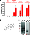
Fig. 5. Reactivity and selectivity analysis of the tetrazene forming reaction on pentanucleotides with an alkylamine in different locations. (A) Bar graph showing fluorescence intensity of 5′-TTTTT-3′ with and without an alkylamine and treated with fluorophore azide 1k. Reaction conditions: KHCO3 (20 mM), 5′-TTTTT-3′ with and without amine group (0.2 mM), azide 1k (3 mM), and PPh3 (3 mM) in BMPy OTf at 50 °C for a specific time. Error bars represent standard deviation (n = 3). Statistical analysis of the data is available in Fig. S9. (B) Chemical structure of alkylazide containing a boron dipyrromethene (BODIPY) group (1k). (C) Agarose gel images for the reaction of TTTTT-5′-NH2 with azide 1k in the presence of DNA ladder (10–300 bps). Total DNA samples were visualized by the fluorescence from SYBR Gold nucleic acid gel stain (Cy3 excitation and emission), while modified DNA samples were visualized by the fluorescence from BODIPY (Cy2 excitation and emission). Reaction conditions: KHCO3 (20 mM), TTTTT with or without NH2 tag at 5′ position (0.2 mM), DNA mixture (0.9 mg mL−1), azide 1k (3 mM), and PPh3 (3 mM) in BMPy OTf at 50 °C for 2 h. Chemical structures of the alkylamine groups on different positions (internal, 3′, and 5′) are available in Fig. S18.
Fig. 6
Fig. 6. Attachment of cholesterol to a DNA aptamer toward the RNA hairpin of human immunodeficiency virus (HIV)-1 transactivation-responsive (TAR) element through the tetrazene-forming reaction. Sequence of the HIV-1-TAR aptamer: 5′-CCCTAGTTAGCCATCTCCC-3′. Modification conditions: HIV-1-TAR-5′-NH2 aptamer (0.1 mM), KHCO3 (20 mM), azide 1l (7.5 mM), and PPh3 (2) or OPPh3 (3) (20 mM) in EMIM OAc/DMF/DMSO (2 : 1 : 1) at 50 °C for 2 h. (A) Chemical structure of cholesterol azide (1l). (B) Agarose gel analysis of HIV-1-TAR-5′-NH2 aptamer modified with cholesterol azide (1l) in the presence of triphenylphosphine oxide (3), triphenylphosphine (2a) JohnPhos (2h), and sulfonate-substituted triphenylphosphine (2e). Degree of conversion was calculated by quantification of the unmodified DNA bands in comparison with the triphenylphosphine oxide condition as 100% of the starting material. (C) Matrix-assisted laser desorption/ionization (MALDI-MS) analysis of the modification of HIV-1-TAR-5′ NH2 aptamer with azide 1l and triphenylphosphine oxide (top, negative control) or triphenylphosphine (bottom). (D) Confocal microscopy images of HeLa cells stained with HIV-1-TAR aptamer hybridized with its complementary DNA-Cy5 conjugate (magenta). Top: cholesterol-modified aptamer (azide/PPh3-treated aptamer). Bottom: unmodified aptamer (azide/OPPh3-treated aptamer). Green: actin filament stain with phalloidin–CF488 conjugate. Scale bar: 20 μm.
Fig. 7
Fig. 7. Tetrazene-forming reaction of DNA aptamer with the Her2 receptor. Sequence of the Her2 aptamer: 5′-GCAGCGGTGTGGGGGCAGCGGTGTGGGGGCAGCGGTGTGGGG-3′. Modification conditions: Her2 aptamer-5′-NH2 aptamer (0.1 mM), KHCO3 (20 mM), biotin azide (7.5 mM), and PPh3 (20 mM) in EMIM OAc/DMF/DMSO (2 : 1 : 1 ratio) at 50 °C for 2 h. (A) Partial crystal structure of Her2 receptor (PDB ID: 1N8Z). (B) Confocal microscopy images of Her2-overexpressing SK-BR-3 cells stained with biotin-modified Her2 aptamer (top), unmodified aptamer (middle), or biotin-modified Her2 aptamer hybridized with complementary DNA sequence (bottom). The bound aptamer was visualized by streptavidin–fluorophore (Cy5) conjugate (1:50 dilution) shown in magenta. Blue: nuclear stain with DAPI. DIC: differential interference contrast. Scale bar: 20 μm.

Similar articles

References

    1. Zheng J. Yang R. Shi M. Wu C. Fang X. Li Y. Li J. Tan W. Chem. Soc. Rev. 2015;44:3036–3055. doi: 10.1039/C5CS00020C. - DOI - PMC - PubMed
    1. Monroy-Contreras R. Vaca L. J. Nucleic Acids. 2011;2011:741723. - PMC - PubMed
    1. Smith C. J. Osborn A. M. FEMS Microbiol. Ecol. 2009;67:6–20. doi: 10.1111/j.1574-6941.2008.00629.x. - DOI - PubMed
    1. Ting R. Lermer L. Perrin D. M. J. Am. Chem. Soc. 2004;126:12720–12721. doi: 10.1021/ja046964y. - DOI - PubMed
    1. Wang X. Feng M. Xiao L. Tong A. Xiang Y. ACS Chem. Biol. 2016;11:444–451. doi: 10.1021/acschembio.5b00867. - DOI - PubMed