. 2022 Feb;12(1):24-36.
doi: 10.21037/cdt-21-490.

Triggering receptor expressed on myeloid cells-2 promotes survival of cardiomyocytes after myocardial ischemic injury through PI3K/AKT pathway

Affiliations

Triggering receptor expressed on myeloid cells-2 promotes survival of cardiomyocytes after myocardial ischemic injury through PI3K/AKT pathway

Cong Fu et al. Cardiovasc Diagn Ther. 2022 Feb.

Abstract

Background: Previous studies have already revealed that triggering receptor expressed on myeloid cells-2 (TREM2) plays a significant protective role during the pathogenesis of ischemia injury in both brain and liver. This study aims to investigate the effect of TREM2 in myocardial ischemic injury.

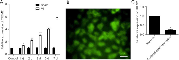
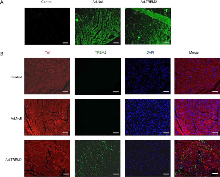
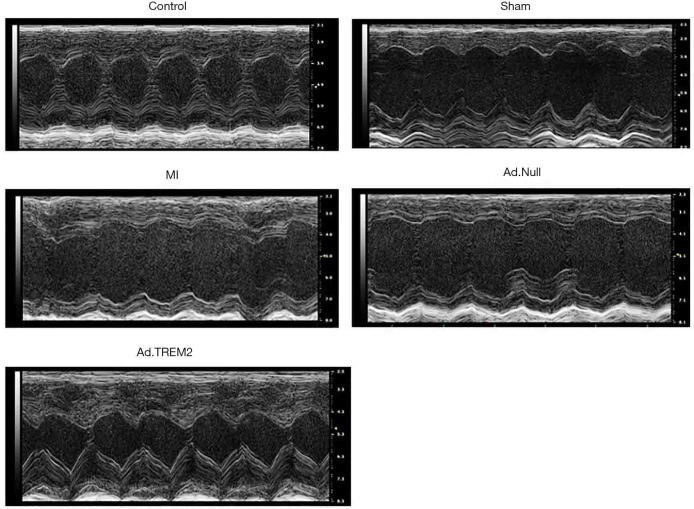
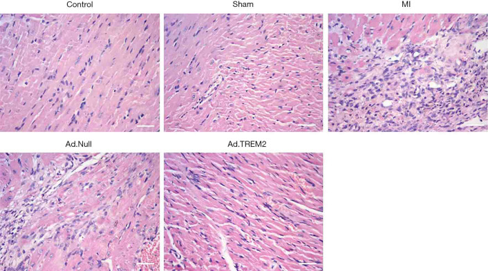
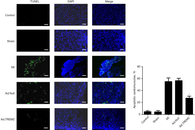
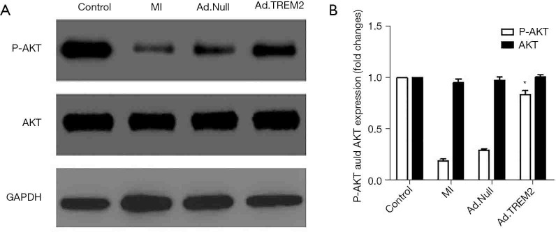
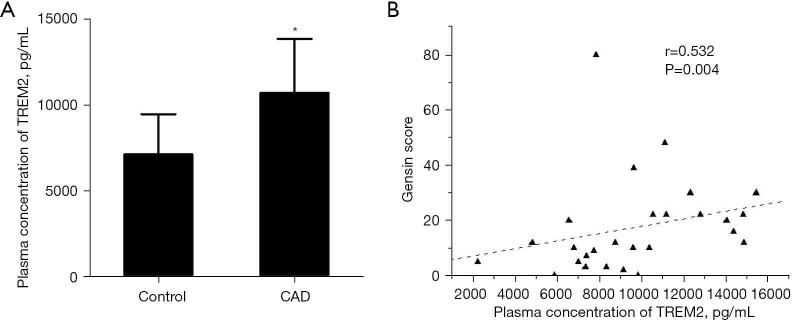
Methods: The mice myocardial infarction (MI) model was established via left anterior descending coronary artery ligation. TREM2 expression was examined with RT-PCR and Western blot. Whereafter, mice were randomly divided into control, sham, MI, Ad.TREM2 transfection group and Ad.Null transfection group. Recombinant adenovirus containing the gene coding full-length mouse TREM2 and EGFP (Ad.TREM2) or control vector containing EGFP gene only (Ad.Null) were immediately intramyocardial injected after left anterior descending ligated. After 7 days of MI, HE, Masson and TUNEL staining were performed to find the myocardial injury, infarcted size and cell apoptosis. Besides, echocardiography was performed to determine cardiac function. In addition, Western blot was performed to check the activity of PI3K/AKT signaling pathway in myocardial tissue. Furthermore, the plasma concentrations of TREM2 in 19 coronary artery disease (CAD) patients and 8 healthy controls were measured.

Results: Compared with the sham group, TREM2 expression was significantly up-regulated in cardiac tissue in mice with MI. Cardiac tissue in mice transfected with Ad.TREM2 was demonstrated with alleviated injury, reduced infarct size, and decreased number of apoptotic cells. Echocardiography revealed that heart function was significantly improved in Ad.TREM2 transfection mice. Also, TREM2 transfection significantly activated the phosphorylation of AKT. At last, the plasma concentration of TREM2 was significantly elevated in patients with CAD and correlated with the severity of CAD.

Conclusions: TREM2 may curb myocardial ischemia injury via activating PI3K/AKT signal pathway. Besides, plasma TREM2 may be treated as a potential biomarker in the diagnosis of CAD to reflect the severity of coronary stenosis.

Keywords: Triggering receptor expressed on myeloid cells-2 (TREM2); apoptosis; cardiac function; myocardial ischemic injury.


Conflict of interest statement

Conflicts of Interest: All authors have completed the ICMJE uniform disclosure form (available at https://cdt.amegroups.com/article/view/10.21037/cdt-21-490/coif). The authors have no conflicts of interest to declare.

Figures

Figure 1
Figure 1
The expression of TREM2 mRNA in myocardial tissue was increased after MI and cultured neonatal mouse cardiomyocytes lowly expressed TREM2. (A) RT-PCR showed that after 1 d, 2 d, 3 d, 5 d and 7 d of MI, the expression of TREM2 mRNA in myocardial tissue gradually increased (n=5, *, P<0.05 vs. control; **, P<0.05 vs. control, 1 d, 3 d, 5 d and 7 d; ***, P<0.05 vs. control, 1 d, 2 d, 5 d and 7 d; ****, P<0.05 vs. control, 1 d, 2 d, 3 d and 7 d). The sham group showed no increased expression of TREM2. (B) Immunofluorescence showed that cultured neonatal mouse expressed TnI. (C) RT-PCR showed that cultured neonatal mouse cardiomyocytes lowly expressed TREM2 compared to mice BM cells. Magnification: ×400; bar: 50 µm; n=5, *, P<0.05 vs. BM cells. TREM2, triggering receptor expressed on myeloid cells-2; MI, myocardial infarction; RT-PCR, reverse transcription-polymerase chain reaction; BM, bone marrow.
Figure 2
Figure 2
Transfection of TREM2 adenovirus in the heart tissue of mice. After Ad.TREM2 transfection, myocardial tissue expressed GFP and TREM2. (A) Immunofluorescence showed that TREM2 adenovirus and control adenovirus successfully transfected in heart tissue; (B) immunofluorescence showed that heart tissue expressed TREM2 after TREM2 adenovirus transfection. The control and control adenovirus transfection had no TREM2 expression. Magnification: ×200; bar: 100 µm. TREM2, triggering receptor expressed on myeloid cells-2; GFP, green fluorescent protein. DAPI, 4',6-diamidino-2-phenylindole; TnI, troponin I.
Figure 3
Figure 3
Effect of TREM2 on cardiac function after MI. Ad.TREM2 transfection increased cardiac function after myocardial infaction. The impairment of cardiac function occurred in the MI group. The expression of TREM2 prevented cardiac dysfunction after MI. The LVEF and LVFS were significantly higher in the TREM2 transfection group (n=5). TREM2, triggering receptor expressed on myeloid cells-2; MI, myocardial infarction; LVEF, left ventricular ejection fraction; LVFS, left ventricular fractional shortening.
Figure 4
Figure 4
Effect of TREM2 expression on pathological change in myocardial tissue. Ad.TREM2 transfection alleviated myocardial injury after MI. HE staining showed that in the MI group, HE staining showed myocardial dissolving and numbers of inflammatory cells infiltration. TREM2 expression alleviated the myocardial injury after the MI group compared to MI and Ad.Null. Magnification: ×400; bar: 50 µm; n=5. TREM2, triggering receptor expressed on myeloid cells-2; MI, myocardial infarction.
Figure 5
Figure 5
Effect of TREM2 expression on infarcted size after MI. The Ad.TREM2 transfection alleviated myocardial injury after MI, Masson staining showed that TREM2 adenovirus transfection significantly reduced the infarcted size after MI compared to Ad.Null transfection and MI group. Histogram showed the infarcted size in all groups. Magnification: 4×; n=5, *, P<0.05 vs. MI and Ad.Null group. TREM2, triggering receptor expressed on myeloid cells-2; MI, myocardial infarction.
Figure 6
Figure 6
Effect of TREM2 on cardiomyocyte apoptosis after MI. The Ad.TREM2 transfection decreased cell apoptosis after myocardial infection. TUNEL staining showed that TREM2 adenovirus transfection significantly reduced the number of apoptotic cells after MI compared to Ad.Null transfection and MI group. Histogram showed the number of apoptotic cells in all groups. Magnification: ×200; bar: 100 µm; n=5, *, P<0.05 vs. MI and Ad.Null group. TREM2, triggering receptor expressed on myeloid cells-2; MI, myocardial infarction. DAPI, 4',6-diamidino-2-phenylindole; TnI, troponin I.
Figure 7
Figure 7
Effect of TREM2 expression on the expression of signal protein expression in heart tissue. The Ad.TREM2 transfection activated the phosphorylation of AKT. (A) Representative band showed that in MI, phosphorylation of AKT was inhibited and TREM2 expression increased the expression of phosphorylated AKT. TREM2 promoted the activation of phosphorylation of AKT compared to MI and Ad.Null. (B) Densitometry quantitation of protein expression levels are shown as fold changes in the histogram. n=5, *, P<0.05 vs. MI and Ad.Null group. TREM2, triggering receptor expressed on myeloid cells-2; MI, myocardial infarction. GAPDH, glyceraldehyde-3-phos-phate dehydrogenase.
Figure 8
Figure 8
The plasma concentration of TREM2 in CAD patients and controls. The plasma concentration of TREM2 in CAD patients was significantly higher than controls. (A) Plasma concentration of TREM2 were elevated in CAD patients (*, P=0.008 vs. control); (B) plasma concentration of TREM2 were correlated with severity coronary stenosis (r=0.532, P=0.004). TREM2, triggering receptor expressed on myeloid cells-2; CAD, coronary artery disease.

Similar articles

Cited by

References

    1. Anderson JL, Morrow DA. Acute Myocardial Infarction. N Engl J Med 2017;376:2053-64. 10.1056/NEJMra1606915 - DOI - PubMed
    1. Ritsinger V, Nyström T, Saleh N, et al. Heart failure is a common complication after acute myocardial infarction in patients with diabetes: A nationwide study in the SWEDEHEART registry. Eur J Prev Cardiol 2020;27:1890-901. 10.1177/2047487319901063 - DOI - PubMed
    1. Daws MR, Lanier LL, Seaman WE, et al. Cloning and characterization of a novel mouse myeloid DAP12-associated receptor family. Eur J Immunol 2001;31:783-91. 10.1002/1521-4141(200103)31:3<783::AID-IMMU783>3.0.CO;2-U - DOI - PubMed
    1. Colonna M. TREMs in the immune system and beyond. Nat Rev Immunol 2003;3:445-53. 10.1038/nri1106 - DOI - PubMed
    1. Ford JW, McVicar DW. TREM and TREM-like receptors in inflammation and disease. Curr Opin Immunol 2009;21:38-46. 10.1016/j.coi.2009.01.009 - DOI - PMC - PubMed