. 2022 Mar 23;144(11):4925-4941.
doi: 10.1021/jacs.1c12745. Epub 2022 Mar 12.

Selective Recognition of Carbohydrate Antigens by Germline Antibodies Isolated from AID Knockout Mice

Affiliations

Selective Recognition of Carbohydrate Antigens by Germline Antibodies Isolated from AID Knockout Mice

Andrew T DeLaitsch et al. J Am Chem Soc. .

Abstract

Germline antibodies, the initial set of antibodies produced by the immune system, are critical for host defense, and information about their binding properties can be useful for designing vaccines, understanding the origins of autoantibodies, and developing monoclonal antibodies. Numerous studies have found that germline antibodies are polyreactive with malleable, flexible binding pockets. While insightful, it remains unclear how broadly this model applies, as there are many families of antibodies that have not yet been studied. In addition, the methods used to obtain germline antibodies typically rely on assumptions and do not work well for many antibodies. Herein, we present a distinct approach for isolating germline antibodies that involves immunizing activation-induced cytidine deaminase (AID) knockout mice. This strategy amplifies antigen-specific B cells, but somatic hypermutation does not occur because AID is absent. Using synthetic haptens, glycoproteins, and whole cells, we obtained germline antibodies to an assortment of clinically important tumor-associated carbohydrate antigens, including Lewis Y, the Tn antigen, sialyl Lewis C, and Lewis X (CD15/SSEA-1). Through glycan microarray profiling and cell binding, we demonstrate that all but one of these germline antibodies had high selectivity for their glycan targets. Using molecular dynamics simulations, we provide insights into the structural basis of glycan recognition. The results have important implications for designing carbohydrate-based vaccines, developing anti-glycan monoclonal antibodies, and understanding antibody evolution within the immune system.


Conflict of interest statement

The authors declare no competing financial interest.

Figures

Figure 1.
Figure 1.
Assembly of antibody genes. Antibodies are composed of two light chains and two heavy chains. The sequences for these chains are assembled via recombination of V, D, and J genes for the heavy chain and V and J genes for the light chain. The D genes are relatively short and can be trimmed, often resulting in lengths of only 5–15 nucleotides. Diversity is further increased by insertion of nontemplated nucleotides in the V–D and D–J junctions of the heavy chain.
Figure 2.
Figure 2.
Structures of selected Lewis Y and related conjugates. (A) Lewis Y hapten conjugates were prepared via reductive amination to free amines on albumin (lysine side chains and/or the N-terminus). (B) Glycans and linkers for selected array components.
Figure 3.
Figure 3.
Comparison of anti-Lewis Y antibody sequences. (A) Amino acid sequence alignment of anti-Lewis Y antibodies using 8F11 as the reference sequence. (B) Nucleotide and amino acid sequence of the VDJ junction region of 8F11 compared to H18A/H18A inferred germline (H18A-iGL). The sequence of H18A is identical to the inferred germline for the nucleotides shown. Junctional nucleotides for H18A-iGL are shown in red. Germline V and J gene assignments for 8F11/H18A are provided. IMGT complementary determining regions are shown in color.
Figure 4.
Figure 4.
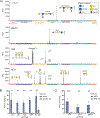
Selective recognition of Lewis Y by germline antibodies. (A) Bar graph showing signals in relative fluorescence units (RFUs) for antibody 8F11 on our 738-component array. Data are shown at a concentration approximately 10-fold higher than the apparent KD value for LeY-tetra to highlight any potential cross-reactivity. Positive and negative controls have been omitted for clarity. Structures are depicted for glycans with signals over 2000 RFU. LeY tetra and LeY-08 have different linkers—see Figure 1. (B) Direct comparison of germline anti-Lewis Y antibody binding to a subset of Lewis Y related glycans. (C) Binding of 8F11 to the Lewis Y positive MCF7 cell line (left) and to the Lewis Y negative SK-MEL-28 cell line (right) as compared to an IgM isotype control. Bar graphs for 12D6 and 8G6 can be found in Figure S3. Additional flow cytometry data can be found in Figures S4 and S5.
Figure 5.
Figure 5.
Binding properties of representative germline antibodies to Lewis X, sialyl Lewis C, and the Tn antigen. (A) Signals in relative fluorescence units (RFUs) for antibodies on our 738-component array. Data for 11A11, 9A9, and 8C11 are at a concentration approximately 20-fold higher than KD,App for the primary antigen; 2D3.C11 is at 8 μg/mL. Positive and negative controls have been excluded. (B) Binding data for anti-Lewis X antibodies to Lewis X positive MCF7 cell line and Lewis X negative SK-MEL-28 as compared to positive control anti-Lewis X antibody MC-480. (C) Binding data for anti-Tn antibodies to Tn positive MCF7 cell line and Tn negative HEK-293 cell line as compared to positive control anti-Tn antibody SBH-Tn. Error bars (B,C) represent the SD of three experiments. Additional bar graphs can be found in Figure S8. Representative histograms and additional flow cytometry data can be found in Figure S9.
Figure 6.
Figure 6.
Structures of Lewis X (A), sialyl Lewis C (B), and Tn-related (C) array components.
Figure 7.
Figure 7.
Modeled structures of antibodies 8F11 and H18A. (A–D) Worm plots illustrating the average magnitude of the RMS atomic fluctuation in each backbone atom over the course of the simulation mapped to the radius of the backbone trace. Regions of the structure that are relatively stable and rigid appear narrower, and more flexible regions of the structure appear wider. View looking down into the binding pocket with the heavy chain on the left and the light chain on the right. CDR loops are labeled, and residues that differ between germline and affinity mature are shown in stick representation.
Figure 8.
Figure 8.
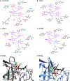
Interactions between Lewis Y and antibodies. (A–D) Schematic view of the hydrogen bond networks. (A) H-bonding for hu3S193 was determined from the crystal structure; (B–D) H-bonding shown was present at least 10% of the time in the molecular dynamics simulations. (E,F) Cluster representative snapshots illustrating residue differences between antibodies. (E) 8F11 germline, with residue D109 in the CDRH3 loop colored green. (F) H18A affinity mature, with residue Y109 colored green. Hydrogen bonds are shown as orange lines.
Figure 9.
Figure 9.
Worm plots, snapshot, and electrostatic surface potential of 12D6. (A,B) Worm plots illustrating the average magnitude of the RMS atomic fluctuation in each backbone atom over the course of the simulation mapped to the radius of the backbone trace. Regions of the structure that are relatively stable and rigid appear narrower, and more flexible regions of the structure appear wider. View looking down into the binding pocket with the heavy chain on the left and the light chain on the right. CDR loops are labeled, and residues that differ between germline and affinity mature are shown in stick representation. (C) Cluster representative snapshot. Hydrogen bonds are shown as orange lines. (D) Top-down view of the antigen binding site surface in 12D6, colored by electrostatic potential. Lewis Y is shown in the pocket, with the linker attachment point marked with a violet ball. The linker is in close proximity to residues D109 and Y110 of the heavy chain (IMGT numbering).

Similar articles

Cited by

References

    1. Schroeder HW Jr.; Cavacini L Structure and function of immunoglobulins. J. Allergy Clin. Immunol. 2010, 125, S41–S52. - PMC - PubMed
    1. Leavy O Therapeutic antibodies: Past, present and future. Nat. Rev. Immunol. 2010, 10, 297. - PubMed
    1. Vajda S; Porter KA; Kozakov D Progress toward improved understanding of antibody maturation. Curr. Opin. Struct. Biol. 2021, 67, 226–231. - PMC - PubMed
    1. Jimenez R; Salazar G; Yin J; Joo T; Romesberg FE Protein dynamics and the immunological evolution of molecular recognition. Proc. Natl. Acad. Sci. U.S.A. 2004, 101, 3803–3808. - PMC - PubMed
    1. Nguyen HP; Seto NOL; MacKenzie CR; Brade L; Kosma P; Brade H; Evans SV Germline antibody recognition of distinct carbohydrate epitopes. Nat. Struct. Biol. 2003, 10, 1019–1025. - PubMed

Publication types

LinkOut - more resources