. 2022 Jun:213:102264.
doi: 10.1016/j.pneurobio.2022.102264. Epub 2022 Mar 11.

Ultrastructural view of astrocyte arborization, astrocyte-astrocyte and astrocyte-synapse contacts, intracellular vesicle-like structures, and mitochondrial network

Affiliations

Ultrastructural view of astrocyte arborization, astrocyte-astrocyte and astrocyte-synapse contacts, intracellular vesicle-like structures, and mitochondrial network

Sydney Aten et al. Prog Neurobiol. 2022 Jun.

Abstract

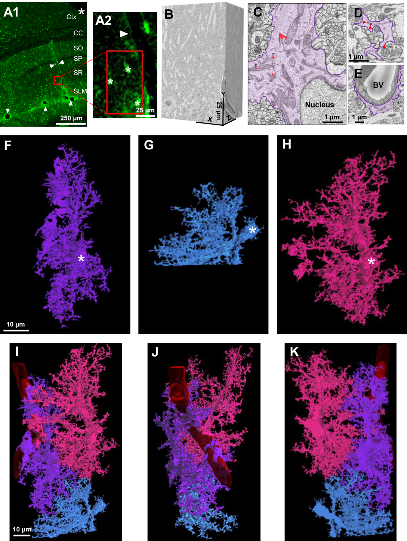
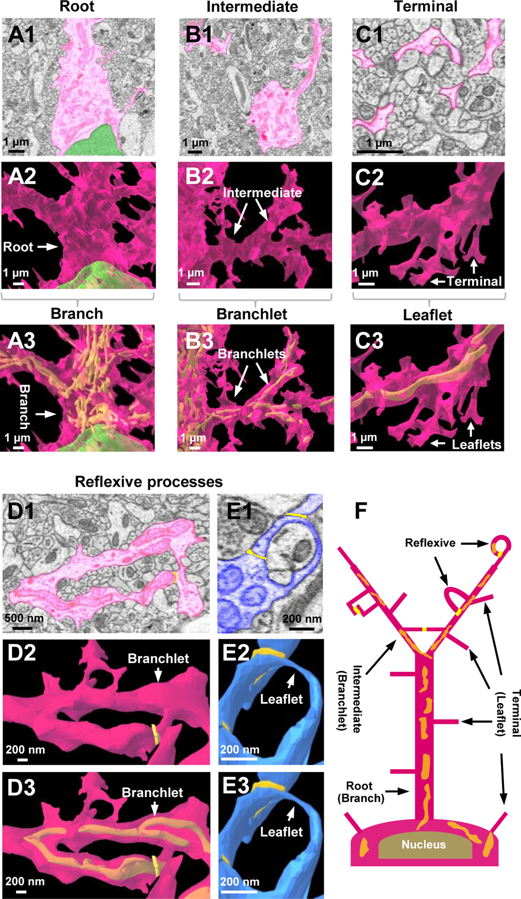
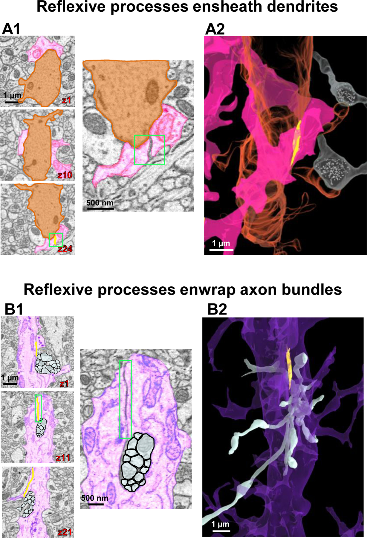
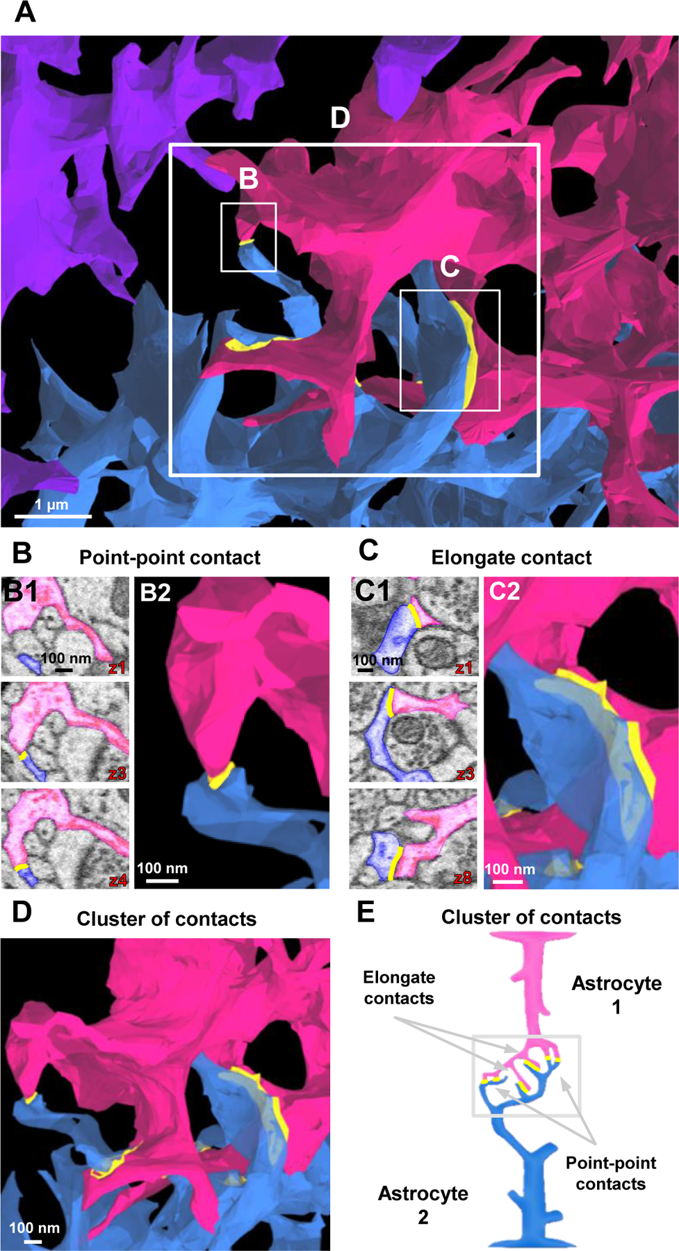
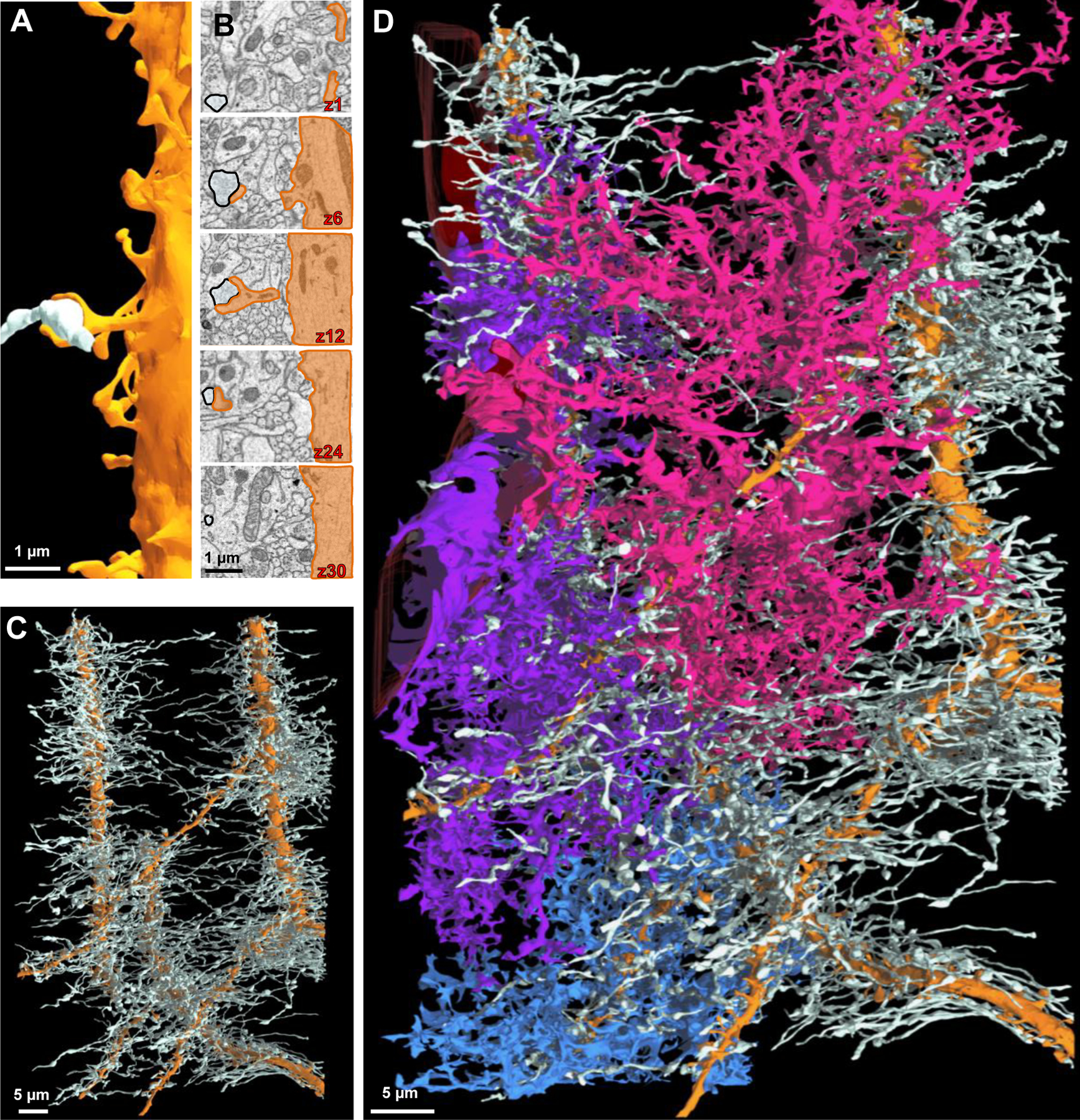
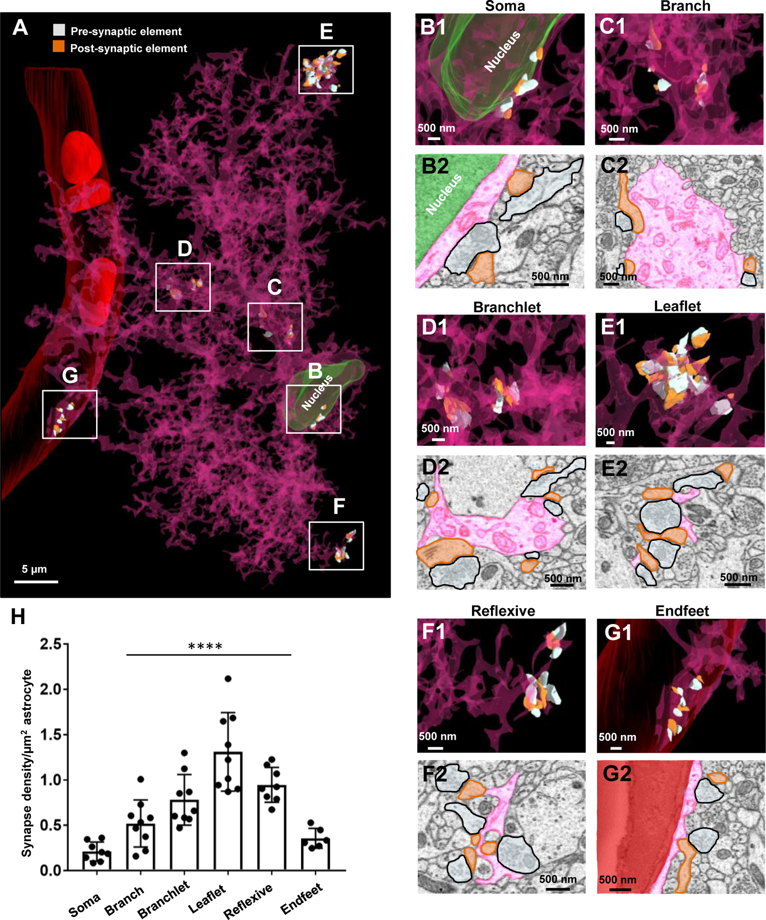
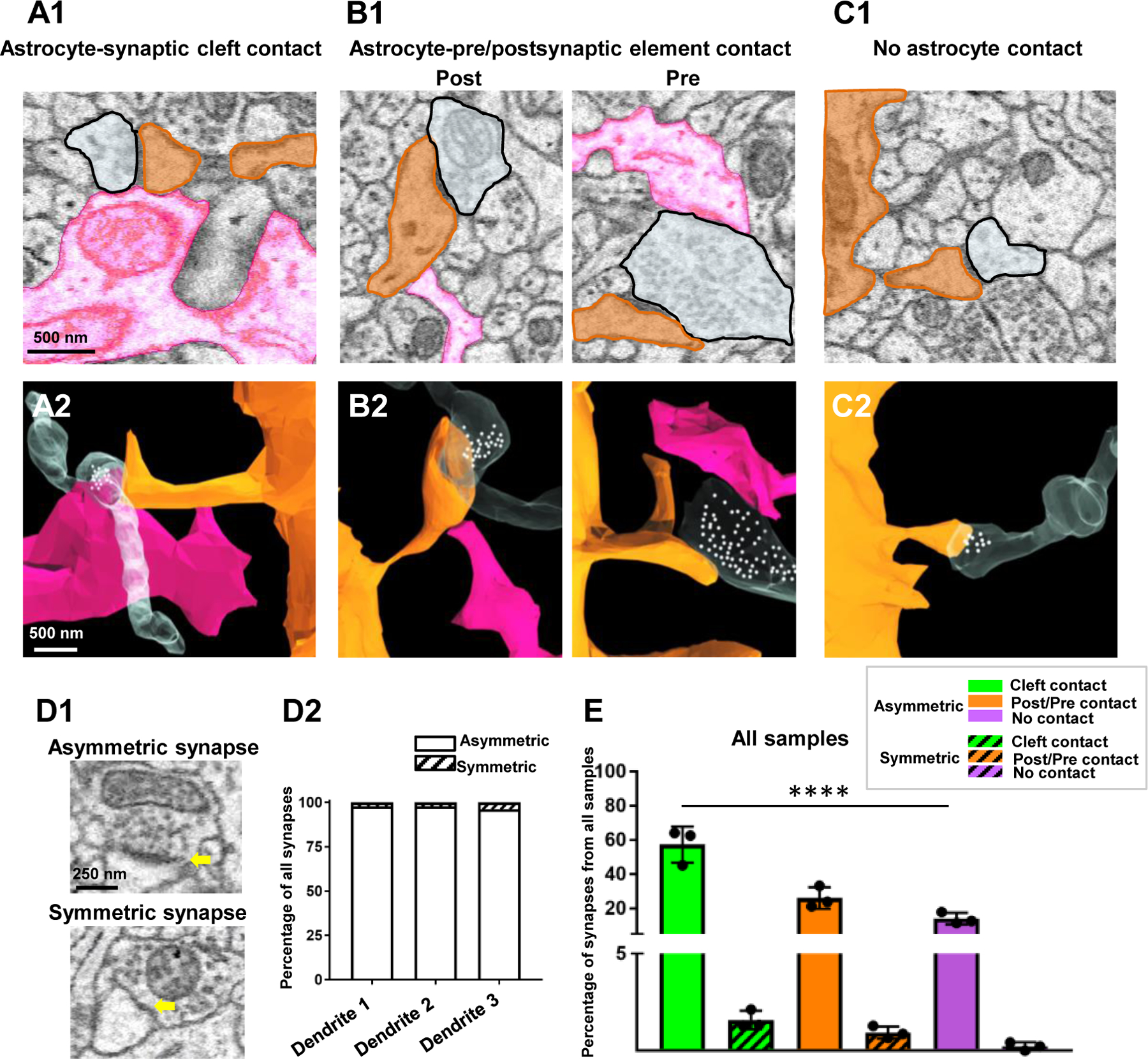
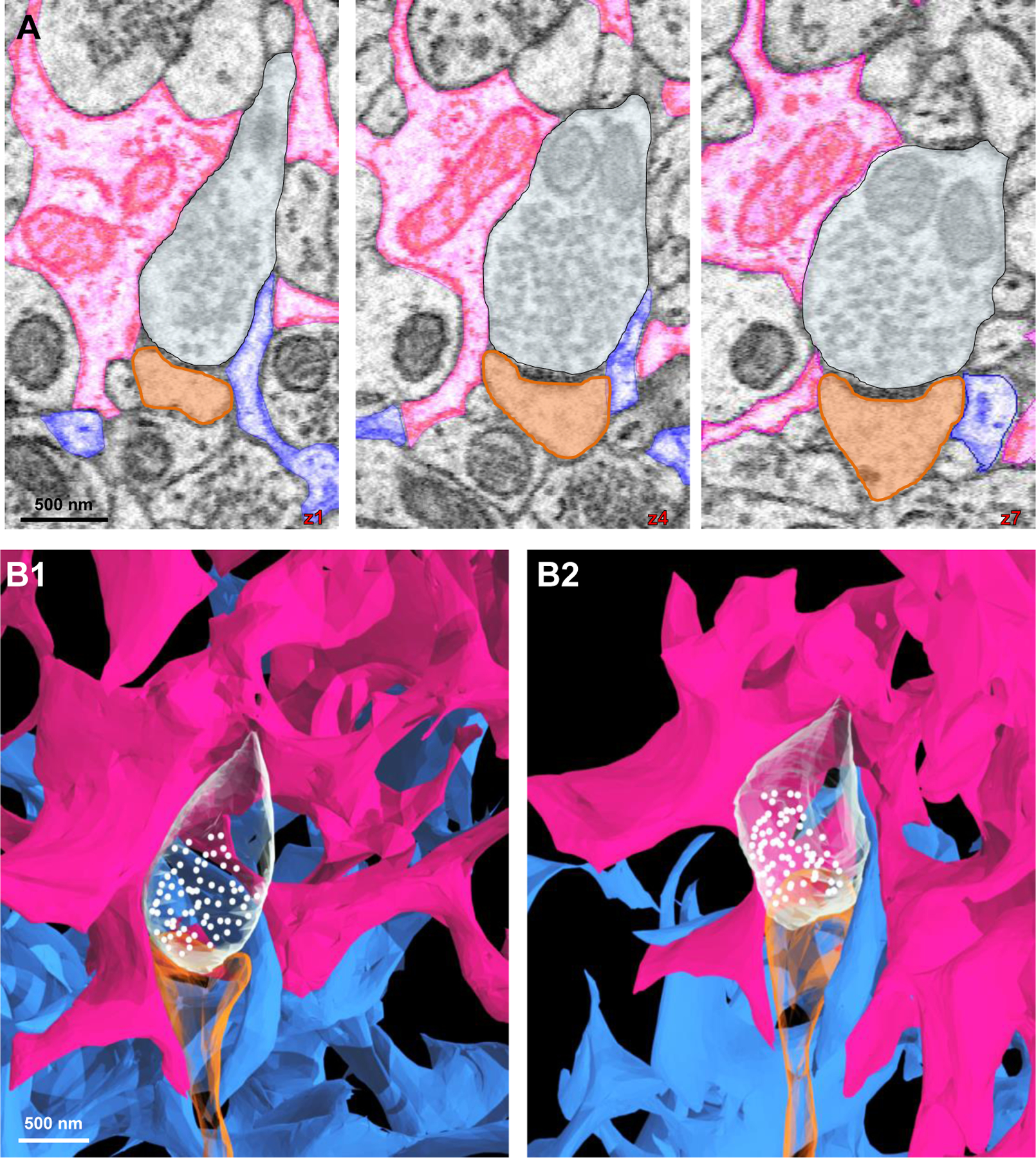
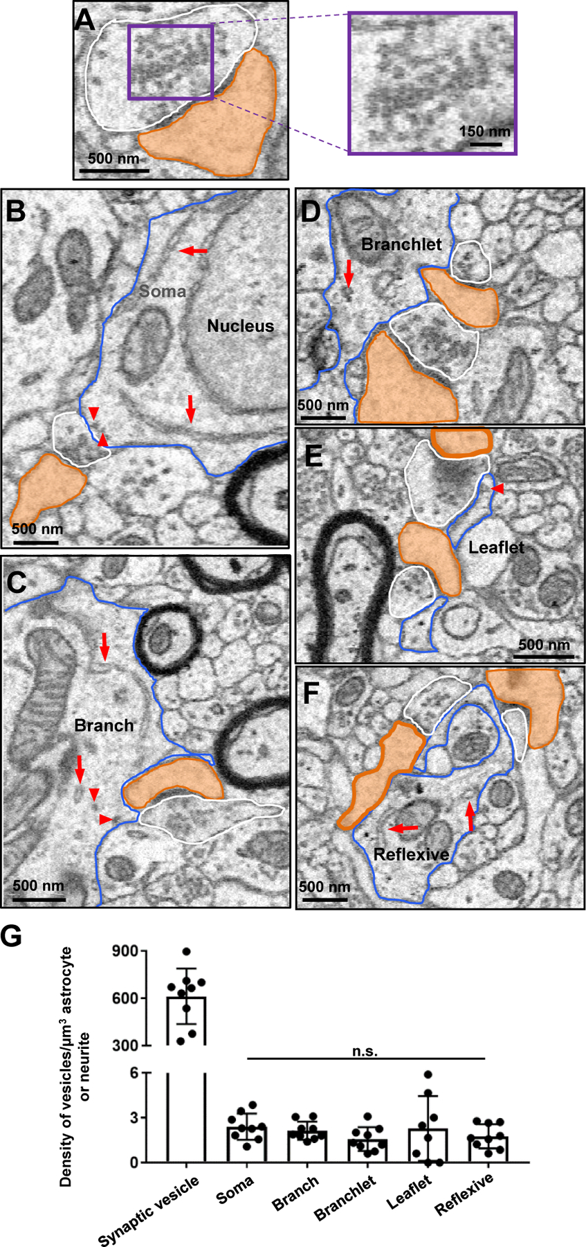
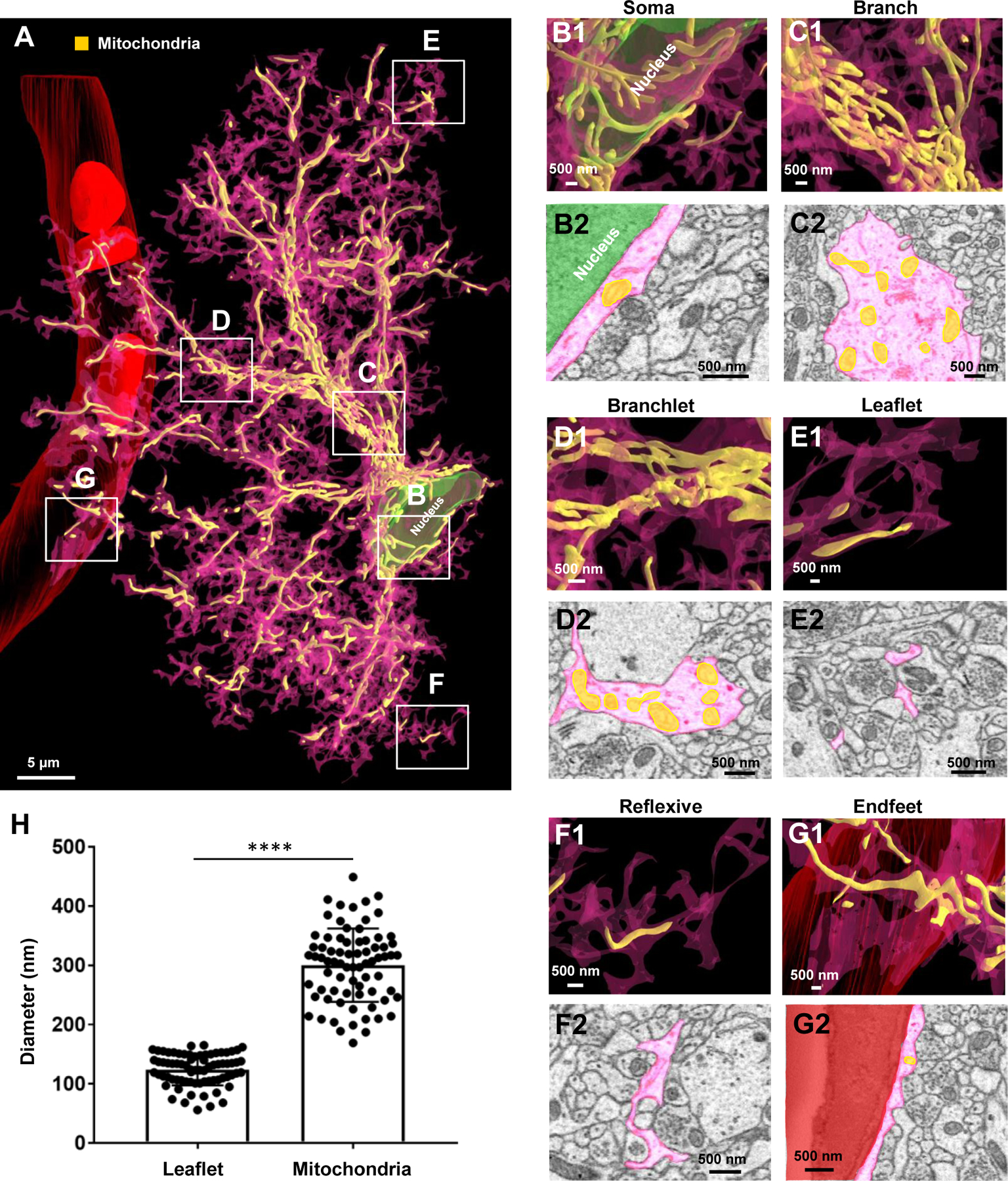
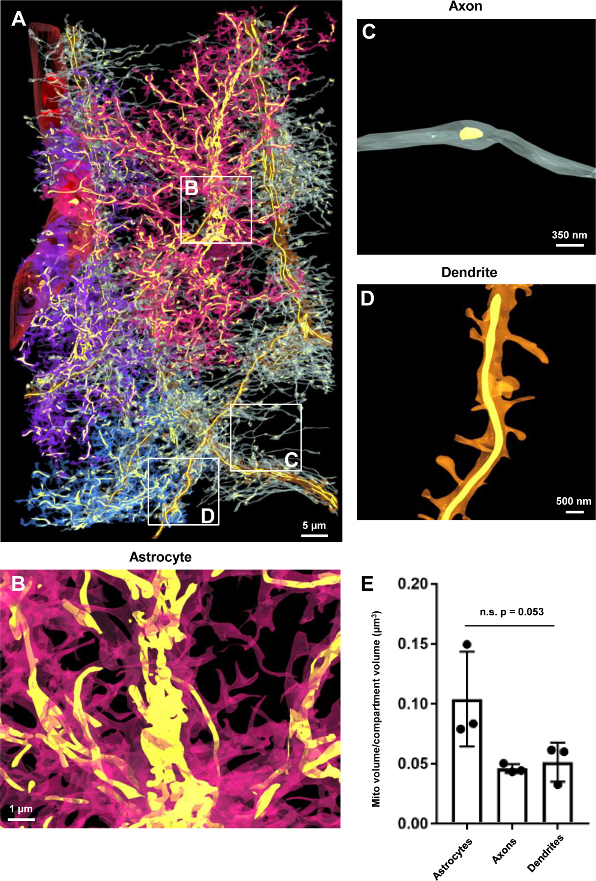
The complexity of astrocyte morphology and syncytial coupling through gap junctions are crucial for astrocyte function in the brain. However, the ultrastructural details of astrocyte arborization and interactions between neighboring astrocytes remain unknown. While a prevailing view is that synapses selectively contact peripheral astrocyte processes, the precise spatial-location selectivity of synapses abutting astrocytes is unresolved. Additionally, knowing the location and quantity of vesicles and mitochondria are prerequisites to answer two emerging questions - whether astrocytes have a signaling role within the brain and whether astrocytes are highly metabolically active. Here, we provided structural context for these questions by tracing and 3D reconstructing three neighboring astrocytes using serial block-face scanning electron microscopy. Our reconstructions reveal a spongiform astrocytic morphology resulting from the abundance of reflexive and leaflet processes. At the interfaces, varying sizes of astrocyte-astrocyte contacts were identified. Inside an astrocyte domain, synapses contact the entire astrocyte, and synapse-astrocyte contacts increase from soma to terminal leaflets. In contrast to densely packed vesicles at synaptic boutons, vesicle-like structures were scant within astrocytes. Lastly, astrocytes contain dense mitochondrial networks with a mitochondrial volume ratio similar to that of neurites. Together, these ultrastructural details should expand our understanding of functional astrocyte-astrocyte and astrocyte-neuron interactions.

Keywords: Aldh1l1-eGFP; Astrocyte network; Mitochondria; Serial blockface scanning electron microscopy (SBF-SEM); Synapses; Synaptic-like microvesicles; Three-dimensional reconstruction.


Conflict of interest statement

Conflict of Interest Statement:

The authors declare no competing interests.

Figures

Figure 1.
Figure 1.. EM Identification and 3D reconstruction of neighboring astrocytes.
A1) Low magnification confocal microscope image of a fixed brain section displaying the location of eGFP+ astrocytes and delineation of blood vessels (white arrowheads). An angular cut (*) at the upper-right region of the tissue serves as a fiduciary mark. A2) A magnified area from A1 shows the spatial location of 3 neighboring astrocytes next to a blood vessel. Asterisks (*) denote the 3 astrocytes observed in the SBF-SEM images. B) The resulting 500-stack SBF-SEM volume dataset from the selected ROI containing neighboring astrocytes from the stratum radiatum hippocampal subregion. C) An astrocyte was identified by first locating the nucleus-containing cell body. Bundles of filaments (large, red arrow) and several examples of glycogen granules (red arrowheads, also see Fig. S1) are noted near the astrocytic nucleus. D) Astrocyte processes (purple) that extend from the cell body possess an irregular and angular shape. E) Processes that contact blood vessels expand into specialized astrocyte endfeet processes. Abbreviations: Ctx-Cortex, CC-Corpus callosum, SO-Stratum oriens, SP-Stratum pyramidale, SR-stratum radiatum, SLM-stratum lacunosum-moleculare, BV-blood vessel. F-H) 3-dimensional view of three reconstructed astrocytes: purple, blue, and pink. White asterisks found within each cell denote the somas, respectively. I-K) Combined reconstruction depicting the front, side, and back views of the three astrocytes. Each astrocyte is labeled in a different color to clearly demarcate individual astrocyte domains and cellular structures. Note that the blue astrocyte appears ‘smaller’ in size as only part of the cell was included in the EM stack.
Figure 2.
Figure 2.. Ultrastructural view of astrocyte processes.
A1) 2D EM trace of an astrocyte root process. Root processes stem directly from the cell body of the astrocyte. The astrocyte nucleus is labeled in green. A2-A3) Reconstructed 3D view of an astrocyte root extending from the cell body. Similar to images in the top panels, the nucleus is shown in green. Note that the root processes are synonymous with previously defined astrocyte ‘branches’. Also note the mitochondria (in yellow) found within the astrocyte branch. The mitochondria (if present) can be observed in the bottom panel of all reconstructions depicted within this figure. B1) 2D EM trace of an astrocyte intermediate process extending from the root process. B2-B3) Reconstructed 3D view of astrocyte intermediate processes branching out from the root process. The intermediate processes are synonymous with previously defined astrocyte ‘branchlets’. Also note the mitochondria (in yellow) found within the branchlets. C1) 2D EM trace of terminal astrocyte processes. C2-C3) Reconstructed 3D view of astrocyte terminal processes extending from an intermediate process. Note that terminal processes are smaller than root and intermediate processes; they reach a terminal (endpoint) and do not branch any further. These processes are synonymous with previously defined astrocyte ‘leaflets’. Note that no mitochondria is found within the leaflets. D1) 2D EM image and 3D reconstruction (D2-D3) of an astrocyte branchlet leading to a blood vessel. This process branches into two processes, which creates a reflexive contact near the endfoot process. Reflexive contacts are indicated in yellow in both the 2D images in the 3D images. Note that these reflexive contacts are depicted with a bright yellow color, relative to the lighter mitochondria, which are strictly found inside the astrocytes. E1) An EM image and 3D reconstruction (E2-E3) of a single leaflet process looping back to the branchlet. A distal leaflet process makes a reflexive contact near the top of the loop. Note that these reflexive processes are not completely fused (i.e., they do not form closed ‘loop’ structures). Also note that panels E2 and E3 are identical given that no mitochondria can be observed within the leaflets. F) Simplified schematic diagram depicting the astrocyte branching architecture and reflexive processes. Note that the diagram depicts both our astrocyte process terminology and the previously-defined branch, branchlet, leaflet terminology. Presumed contacts are noted in bright yellow, while internal mitochondria appears orange (given its location inside the pink astrocyte).
Figure 3.
Figure 3.. Astrocyte reflexive processes and their structural support to neurites.
A1) Serial 2D EM images of astrocyte branchlets (pink) encircling a dendritic shaft (orange). The bottom image in the left panel exhibits a reflexive contact (boxed in green). Higher magnification images in the right panel clearly show the contact depicted in z slice 24. A2) 3D reconstruction showing the astrocyte branchlets (pink) wrap around the dendrite (orange). Two axons (white) in synaptic contact with the associated dendritic spines are also represented. B1) 2D serial EM images of a branch point at the end of an astrocyte root process (purple) that splits into two branchlets, which enwrap a bundle of axons (white). Note the astrocyte reflexive contact in the middle panel (magnified on right side). B2) Side view of a 3D reconstruction of the axons (white) protruding through the enwrapment of the purple astrocyte process. All 3D reconstructions throughout the figure originated from the same astrocyte regions depicted in the corresponding 2D EM traces.
Figure 4.
Figure 4.. Contact patterns of astrocytes at their interfaces.
A) 3D reconstruction of astrocyte processes reveals the interface between the three astrocytes. For ease of viewing the ultrastructural inter-astrocyte contacts (between the pink and blue astrocytes), fewer intermediate and terminal processes were reconstructed in this image (relative to the images depicted in Fig. 1). Also note that contacts boxed in white are magnified in B-D. B) 2D serial EM images (B1) of point-point contacts between two astrocytes. These point-point contacts persist through a few serial sections to create a small contact at the tip of the terminal processes (see 3D EM image in B2. C) 2D serial EM images (C1) of an elongate contact. Elongate contacts typically contact/persist through many serial sections to create a large contact area (see 3D EM image in C2. D) A 3D reconstruction of a ‘cluster of contacts’. This cluster of contacts contains a combination of both point-point and elongate contact types. E) Schematic diagram illustrating point-point contacts and elongate contacts that ultimately form a ‘cluster of contacts’. Throughout the figure, note that all 2D traces are from the same astrocyte region used to generate the 3D reconstructed images.
Figure 5.
Figure 5.. An ultrastructural view of astrocyte-neurite association.
A) Partial 3D reconstruction of one dendrite. An axon (white) is drawn for reference in order to depict the axon-dendritic spine interface (i.e., synapse). B) 2D serial traces of the axon (white) and dendritic spine (orange) that form a synapse (synapse is depicted in A and in z-section 12). C) 3D reconstruction of three dendrites (orange) and their associated axons (white) shown in a front view. D) Front view of the three neighboring astrocytes and their association(s) with the three reconstructed neurites.
Figure 6.
Figure 6.. Synapses contact each compartment of an astrocyte, though an enrichment of synapses abut astrocytic leaflet and reflexive processes.
A) 3D reconstruction of an entire astrocyte (pink) and its contacts with synapses (orange: postsynapse and white: presynapse). Note that the white, boxed areas in A approximate the locations of magnified images in B-G. Further, the synapses depicted in the representative image (A) for each region were constructed from approximately the same volume of astrocyte. B1) Magnified 3D reconstruction of multiple synapses contacting the astrocyte soma. B2) 2D EM trace of synapses contacting the astrocyte soma. Note that the astrocyte nucleus is depicted in green in both the 2D and 3D images. C1) 3D reconstruction and 2D trace (C2) of synapses contacting an astrocyte branch. D1) 3D reconstruction and 2D trace (D2) of synapses contacting astrocyte branchlets. E1) 3D reconstruction and 2D trace (E2) of synapses contacting astrocyte leaflets. F1) 3D reconstruction and 2D trace (F2) of synapses contacting reflexive astrocyte processes. G1) 3D reconstruction and 2D trace (G2) of synapses contacting the astrocyte endfeet adjacent to the blood vessel (red). H) Graphical representation of the synapse density per area of astrocyte. Data was obtained from three reconstructed astrocytes, with three ROIs chosen per astrocyte process region (soma, branch, branchlet, leaflet, reflexive, endfeet); represented as mean ± SEM. Note that one of three astrocytes (blue) did not contact a blood vessel, and thus only six ROIs are shown in the ‘endfoot’ panel of the graph. Also note that both branchlet and leaflet processes were included in the ‘reflexive’ analysis. ****: p < 0.0001; One-way ANOVA, followed by post-hoc tests.
Figure 7.
Figure 7.. The majority of synapses are contacted by astrocytic processes.
A1) 2D EM trace of an astrocyte process (pink) contacting the synaptic cleft. A2) 3D reconstruction from A1. B1) 2D EM traces of astrocyte processes contacting either post-synaptic dendritic elements (left panel) or pre-synaptic elements (right panel). B2) 3D reconstruction from B1. C1) 2D EM trace of a synapse with no astrocyte contact. A 3D reconstruction is also depicted in C2. In all representative images, the astrocyte processes that contact the synapses are from one astrocyte (pink) and the synapses are from two fully reconstructed dendrites shown in Fig. 5. White spheres depict the approximate locations of synaptic vesicles observed from several serial 2D EM stacks. D1) 2D EM traces depict an example of an asymmetric synapse (prominent post synaptic density - top panel) and a symmetric synapse (modest post synaptic density - bottom panel). Yellow arrows denote the post-synaptic density. D2) Graphical representation of the percentage of asymmetric versus symmetric synapses (irrespective of astrocyte contact type) from all three traced dendrites. E) Graphical representation of the percentage of synapses (asymmetric or symmetric) that contact astrocyte processes at the synaptic cleft, on pre- or post-synaptic elements, or have no contact with astrocyte processes. Note that these data were pooled from the synapses that contacted spines from all three reconstructed dendrites, and thus, each data point is representative of the percent coverage per dendrite. Data was analyzed by one-way ANOVA followed by post hoc tests; ****: p < 0.0001.
Figure 8.
Figure 8.. Synapses can be ensheathed by processes stemming from different astrocyte domains.
A) 2D serial EM traces of two astrocyte processes (pink and blue) contacting the same synapse (white: axon; orange: dendritic spine head). B1-B2) 3D reconstructions of the two astrocytes contacting the same synapse (from A). Note that as shown in the 2D traces, the pink process contacts the synapse on the left side and the blue process on the right side.
Figure 9.
Figure 9.. Paucity of vesicle-like structures in synapse-ensheathing astrocyte processes.
A) Representative 2D EM image of a synapse (white: pre-synaptic element; orange: dendritic spine head). Note the significant number of vesicles in the pre-synaptic element (magnified in the right panel). B) 2D EM traces of an astrocyte soma (blue outline), (C) astrocyte branch (blue outline), (D) astrocyte branchlet (blue outline), (E) astrocyte leaflet (blue outline), and (F) astrocyte reflexive process (blue outline). Synapses are also outlined (white: presynaptic element; orange: dendritic spine head). Note that very few (if any) presynaptic microvesicle-like structures were found in astrocyte processes that contacted synapses. Likely astrocyte vesicles are denoted by red arrowheads, and endoplasmic reticulum is denoted by long red arrows. G) Quantification of the approximate number of vesicle-like structures in each astrocyte (or neurite). Data is represented as mean ± SEM. No significant difference in the density of vesicle-like structures was found between process types. n.s.: not significant.
Figure 10.
Figure 10.. Extensive mitochondrial networks in astrocyte soma, branch, branchlet, reflexive, and endfeet processes.
A) 3D reconstruction of an entire astrocyte (pink) and its internal mitochondria (yellow). Note that the white, boxed areas in A approximate the locations of magnified images in B-G. B1) Magnified 3D reconstruction of mitochondria found within the astrocyte soma. B2) 2D EM trace of mitochondria within the astrocyte soma. Note that the astrocyte nucleus is depicted in green in both the 2D and 3D images. C1) 3D reconstruction and 2D trace (C2) of mitochondria within an astrocyte branch. D1) 3D reconstruction and 2D trace (D2) of mitochondria within astrocyte branchlets. E1) 3D reconstruction and 2D trace (E2) of mitochondria within astrocyte leaflets. F1) 3D reconstruction and 2D trace (F2) of mitochondria within reflexive astrocyte processes. G1) 3D reconstruction and 2D trace (G2) of mitochondria within an astrocyte endfoot adjacent to the blood vessel (red). H) Graphical representation depicting the average diameter of leaflet astrocyte processes, relative to the diameter of mitochondria. Each dot is indicative of the diameter of one leaflet process or one mitochondria. The diameter of 25 leaflet processes and 25 mitochondria were measured for each of the three astrocytes. ****: p < 0.0001; student’s t-test.
Figure 11.
Figure 11.. Astrocytes contain a similar quantity of mitochondria compared to neurites.
A) 3D reconstruction of the blood vessel, the three astrocytes, the three dendrites, and the associated axons and internal mitochondria (yellow) found within each structure. Note that the astrocytes and neurites were made partially transparent to visualize the mitochondria. Also note the white boxes in A reflect magnified images of mitochondria within an astrocyte (B), an axon (C) and a dendrite (D). E) Graphical representation of the mitochondria to cell volume ratio for astrocytes and neurites. Note that each point on the graph is representative of the mitochondria to volume ratio of the entire structure (i.e., one of three reconstructed astrocytes, dendrites, or all of the axons from one dendrite). n.s.: not significant; One-way ANOVA.

Similar articles

Cited by

References

    1. Aboufares El Alaoui A, Jackson M, Fabri M, de Vivo L, Bellesi M, 2020. Characterization of Subcellular Organelles in Cortical Perisynaptic Astrocytes. Front Cell Neurosci 14, 573944. - PMC - PubMed
    1. Adams SR, Mackey MR, Ramachandra R, Palida Lemieux SF, Steinbach P, Bushong EA, Butko MT, Giepmans BNG, Ellisman MH, Tsien RY, 2016. Multicolor Electron Microscopy for Simultaneous Visualization of Multiple Molecular Species. Cell Chem Biol 23, 1417–1427. - PMC - PubMed
    1. Agarwal A, Wu PH, Hughes EG, Fukaya M, Tischfield MA, Langseth AJ, Wirtz D, Bergles DE, 2017. Transient Opening of the Mitochondrial Permeability Transition Pore Induces Microdomain Calcium Transients in Astrocyte Processes. Neuron 93, 587–605 e587. - PMC - PubMed
    1. Alivisatos AP, Chun M, Church GM, Greenspan RJ, Roukes ML, Yuste R, 2012. The brain activity map project and the challenge of functional connectomics. Neuron 74, 970–74. - PMC - PubMed
    1. Allen NJ, Eroglu C, 2017. Cell Biology of Astrocyte-Synapse Interactions. Neuron 96, 697–708. - PMC - PubMed

Publication types