. 2022 Feb 24:15:829642.
doi: 10.3389/fnmol.2022.829642. eCollection 2022.

Shikonin Attenuates Cochlear Spiral Ganglion Neuron Degeneration by Activating Nrf2-ARE Signaling Pathway

Affiliations

Shikonin Attenuates Cochlear Spiral Ganglion Neuron Degeneration by Activating Nrf2-ARE Signaling Pathway

Hongjie Du et al. Front Mol Neurosci. .

Abstract

The molecular mechanisms that regulate the proliferation and differentiation of inner ear spiral ganglion cells (SGCs) remain largely unknown. Shikonin (a naphthoquinone pigment isolated from the traditional Chinese herbal medicine comfrey root) has anti-oxidation, anti-apoptosis and promoting proliferation and differentiation effects on neural progenitor cells. To study the protective effect of shikonin on auditory nerve damage, we isolated spiral ganglion neuron cells (SGNs) and spiral ganglion Schwann cells (SGSs) that provide nutrients in vitro and pretreated them with shikonin. We found that shikonin can reduce ouabain, a drug that can selectively destroy SGNs and induce auditory nerve damage, caused SGNs proliferation decreased, neurite outgrowth inhibition, cells apoptosis and mitochondrial depolarization. In addition, we found that shikonin can increase the expression of Nrf2 and its downstream molecules HO-1 and NQO1, thereby enhancing the antioxidant capacity of SGNs and SGSs, promoting cells proliferation, and inhibiting cells apoptosis by activating the Nrf2/antioxidant response elements (ARE) signal pathway. However, knockdown of Nrf2 rescued the protective effect of shikonin on SGNs and SGSs damage. In addition, we injected shikonin pretreatment into mouse that ouabain-induced hearing loss and found that shikonin pretreatment has a defensive effect on auditory nerve damage. In summary, the results of this study indicate that shikonin could attenuate the level of oxidative stress in SGNs and SGSs through the Nrf2-ARE signaling pathway activated, induce the proliferation and differentiation of SGNs, and thereby improve the neurological hearing damage in mice. Therefore, shikonin may be a candidate therapeutic drug for endogenous antioxidants that can be used to treat neurological deafness.

Keywords: Nrf2-ARE; auditory nerve damage; ouabain; shikonin; spiral ganglion cells.


Conflict of interest statement

HD was employed by the company Qilu Pharmaceutical Co., Ltd. The remaining authors declare that the research was conducted in the absence of any commercial or financial relationships that could be construed as a potential conflict of interest.

Figures
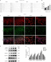
FIGURE 1
FIGURE 1
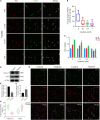
Ouabain damages the hearing of mice and activates SGSs apoptosis and autophagy. (A) Ouabain drugs of different concentrations (0.5, 1.0, and 3 mM) were treated for 7 days, and the auditory brainstem response waveforms of the mice were detected (n = 3). In the control group, the mice were injected with the same dose of saline. (B) Statistical histogram of mouse hearing threshold. (C) Immunofluorescence observed the changes in cell morphology of SGSs explants treated with different concentrations of ouabain (0.1, 0.5, and 1 mM) for 24 h. Red fluorescence represents Tuj1 staining, green fluorescence represents S100 staining, and blue fluorescence represents nuclear (DAPI) staining, bar = 50 μm. (D) After SGNs were treated with different concentrations of ouabain drugs (0.1, 0.5, and 1 mM) for 24 h, cell proteins were collected, and Western Blot was used to detect cell apoptosis and autophagy protein level changes. (E) Statistical histogram of relative protein expression levels. *P < 0.05, **P < 0.01, ***P < 0.001.
FIGURE 2
FIGURE 2
Ouabain affects the differentiation of SGNs into glutamatergic and GABAergic neuronal groups. (A) After separating the SGNs, they were treated with different concentrations of ouabain (0.5, 1.0, and 3 mM) for 24 h, and the cell morphology changes were observed by immunofluorescence. Red represents Tuj1 staining, green fluorescence represents NeuN staining, blue fluorescence represents nuclear (DAPI) staining, bar = 50 μm. (B) Statistics of the average length of SGNs cell body to the apex of the longer synapse after treatment with different concentrations of ouabain. (C) Cell viability (0, 24, and 48 h) after treatment with different concentrations of ouabain was detected by MTT. (D) The expression of VGLUT1 and GAT1 proteins was detected after 24 h of treatment with 0.5 mM ouabain. (E) Statistical histogram of relative expression of VGLUT1 protein. (F) Statistical histogram of the relative expression of GAT1 protein. (G) Immunofluorescence observation of the number of VGLUT1 and GAT1 positive cells after ouabain treatment. Red fluorescence represents Nestin staining, green fluorescence represents VGLUT1 or GAT1 staining, blue fluorescence represents nuclear (DAPI) staining, white arrows represent positive cells, bar = 50 μm. (H) Statistical histogram of the proportion of VGLUT1 and GAT1 positive cells. *P < 0.05, **P < 0.01, ***P < 0.001.
FIGURE 3
FIGURE 3
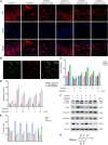
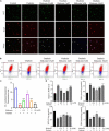
Shikonin can relieve ouabain’s damage to the growth and proliferation of SGNs and SGSs. After pretreatment of SGSs explants with different concentrations of shikonin (0.5, 1, 5, and 10 μM) for 2 h, 0.5 mM ouabain continued treatment for another 22 h. (A) The morphology of SGSs was observed by immunofluorescence. Red fluorescence represents Tuj1 staining, blue fluorescence represents nuclear (DAPI) staining, bar = 50 μm. (B) Nestin and NeuN perform nerve cell labeling. Among them, red represents NeuN staining, green fluorescence represents Nestin staining, and blue fluorescence represents nuclear (DAPI) staining, bar = 50 μm. (C) After treatment with shikonin and ouabain, cell viability detection (0, 24, and 48 h). Flow cytometry was used to detect the changes of apoptosis (D) and cell cycle (E) after treatment with shikonin and ouabain. (F) The effects of shikonin pretreatment on apoptosis and autophagy were detected by Western blot. (G) Structural diagram of shikonin. *P < 0.05, **P < 0.01, ***P < 0.001.
FIGURE 4
FIGURE 4
Shikonin can rescue the effect of ouabain on the differentiation of SGNs. After pretreatment of SGNs with different concentrations of shikonin (0.5, 1, 5, and 10 μM) to separate the cells for 2 h, 0.5 mM ouabain continued treatment for another 22 h. (A) The morphology of SGNs was observed by immunofluorescence. Red represents NeuN staining, green fluorescence represents Nestin staining, blue fluorescence represents nuclear (DAPI) staining, bar = 50 μm. (B) Statistics of the average length of SGNs from the cell body to the apex of the longer synapse in different treatment groups. After collecting cells in different treatment groups, RNA was extracted, and the relative mRNA expression levels of VGLUT1 (C) and GAT1 (D) were detected by qPCR, and β-antin was used as an internal reference gene. (E) The cells of different treatment groups were subjected to protein extraction to detect the expression of VGLUT1 and GAT1 proteins. *P < 0.05, **P < 0.01, ***P < 0.001.
FIGURE 5
FIGURE 5
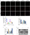
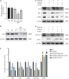
Shikonin can activate the Nrf2/ARE signaling pathway inhibited by ouabain in SGSs. (A) After 0.5 mM ouabain was treated with SGSs explants for different times, cell proteins were collected and the protein expression changes of Nrf2/ARE signaling pathway proteins (Nrf2, HO-1 and NQO1) were detected. (B) Extract the RNA of each group of cells, and detect the relative expression of Nrf2, HO-1 and NQO1 mRNA by qPCR. (C) Immunofluorescence staining to detect the protein expression of Nrf2 after ouabain treatment. Among them, red represents S100 staining, green fluorescence represents Nrf2 staining, blue fluorescence represents nuclear (DAPI) staining, bar = 50 μm. *P < 0.05, **P < 0.01, ***P < 0.001.
FIGURE 6
FIGURE 6
Shikonin can reduce the oxidative stress level of SGNs caused by ouabain. After pretreatment of SGNs with different concentrations of shikonin (0.5, 1, 5, and 10 μM) to separate the cells for 2 h, 0.5 mM ouabain continued treatment for another 22 h. (A) After treatment, the morphology of SGNs cells and the expression of Nrf2 protein were observed. Among them, red represents NeuN staining, green fluorescence represents Nrf2 staining, blue fluorescence represents nuclear (DAPI) staining, bar = 50 μm. (B) The level of mitochondrial depolarization was measured by flow cytometry to detect the JC-1 polymer/monomer fluorescence ratio. Red represents the fluorescence ratio of JC-1 polymer, and blue represents the fluorescence ratio of JC-1 monomer. (C) Different treatment groups, JC-1 red fluorescence/green fluorescence ratio statistical histogram. (D) Detection of secretion levels of oxidative factors ROS and MDA and antioxidant factors SOD and GSH in cells of different treatment groups. *P < 0.05, **P < 0.01, ***P < 0.001.
FIGURE 7
FIGURE 7
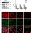
Knockdown of Nrf2 reduces the protective effect of shikonin on SGNs differentiation. (A) The specificity and efficiency of siRNA against Nrf2 were tested by quantitative PCR. (B) The knockout efficiency of Nrf2 siRNA was detected at the protein level by Western blot. SGNs were transfected with Nrf2 siRNA for 24 h, pretreated with shikonin, and then treated with ouabain for another 22 h. To detect the expression changes of Nrf2/ARE signal pathway (Nrf2, HO-1, and NQO1) protein (C) and mRNA (E) in SGNs. As well as the protein (D) and mRNA (E) expression changes of glutamatergic neurons VGLUT1 and GABAergic neurons GAT1. *P < 0.05, **P < 0.01, ***P < 0.001.
FIGURE 8
FIGURE 8
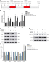
Shikonin has protective effects on ouabain-induced hearing damage in mice. (A) Experimental procedure of drug injection in mice. The arrow represents the drug injection point. The red bars represent the time period of continuous drug injection. The mice in the control group were injected with the same amount of normal saline or drug solvent at the same time point. (B) The statistical histogram of the hearing brainstem response of mice at 7, 14, and 30 days after drug injection (n = 3). After 14 days of drug treatment, detect the expression of Nrf2/ARE signal pathway (Nrf2, HO-1, and NQO1) protein (C) and mRNA (E) in mouse SGN tissue protein. And detect the protein (D) and mRNA (E) expressions of glutamatergic neurons VGLUT1 and GABAergic neurons GAT1. ***P < 0.001.

Similar articles

Cited by

References

    1. Albarakati A. J. A., Baty R. S., Aljoudi A. M., Habotta O. A., Elmahallawy E. K., Kassab R. B., et al. (2020). Luteolin protects against lead acetate-induced nephrotoxicity through antioxidant, anti-inflammatory, anti-apoptotic, and Nrf2/HO-1 signaling pathways. Mol. Biol. Rep. 47 2591–2603. 10.1007/s11033-020-05346-1 - DOI - PubMed
    1. Buchman C. A., Gifford R. H., Haynes D. S., Lenarz T., O’Donoghue G., Adunka O., et al. (2020). Unilateral cochlear implants for severe, profound, or moderate sloping to profound bilateral sensorineural hearing loss: a systematic review and consensus statements. JAMA Otolaryngol. Head Neck Surg. 146 942–953. 10.1001/jamaoto.2020.0998 - DOI - PubMed
    1. Castelnovo L. F., Bonalume V., Melfi S., Ballabio M., Colleoni D., Magnaghi V. (2017). Schwann cell development, maturation and regeneration: a focus on classic and emerging intracellular signaling pathways. Neural Regener. Res. 12 1013–1023. 10.4103/1673-5374.211172 - DOI - PMC - PubMed
    1. Chai R., Li G. L., Wang J., Zou J. (2017). Hearing loss: reestablish the neural plasticity in regenerated spiral ganglion neurons and sensory hair cells. Neural Plast. 2017:1807581. - PMC - PubMed
    1. Chen Y., Gu Y., Li Y., Li G. L., Chai R., Li W., et al. (2021). Generation of mature and functional hair cells by co-expression of Gfi1, Pou4f3, and Atoh1 in the postnatal mouse cochlea. Cell Rep. 35:109016. 10.1016/j.celrep.2021.109016 - DOI - PubMed