Review
. 2022 Feb 25:15:831116.
doi: 10.3389/fnmol.2022.831116. eCollection 2022.

The Unfolded Protein Responses in Health, Aging, and Neurodegeneration: Recent Advances and Future Considerations

Affiliations
Review

The Unfolded Protein Responses in Health, Aging, and Neurodegeneration: Recent Advances and Future Considerations

Andrew P K Wodrich et al. Front Mol Neurosci. .

Abstract

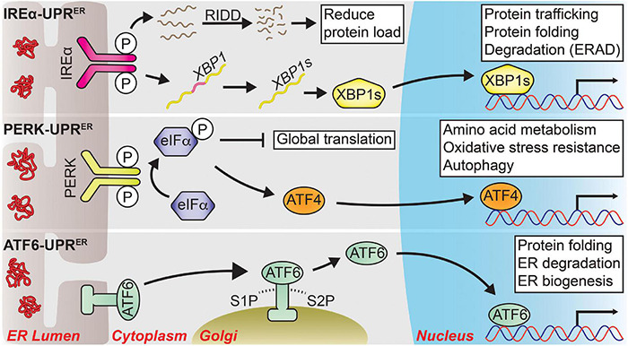
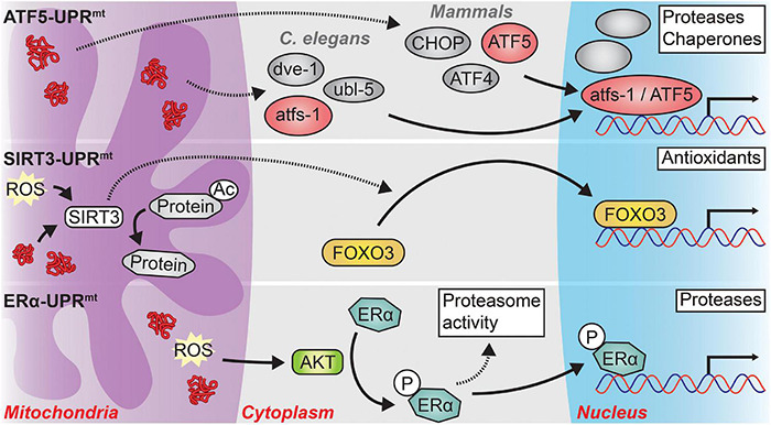
Aging and age-related neurodegeneration are both associated with the accumulation of unfolded and abnormally folded proteins, highlighting the importance of protein homeostasis (termed proteostasis) in maintaining organismal health. To this end, two cellular compartments with essential protein folding functions, the endoplasmic reticulum (ER) and the mitochondria, are equipped with unique protein stress responses, known as the ER unfolded protein response (UPR ER ) and the mitochondrial UPR (UPR mt ), respectively. These organellar UPRs play roles in shaping the cellular responses to proteostatic stress that occurs in aging and age-related neurodegeneration. The loss of adaptive UPR ER and UPR mt signaling potency with age contributes to a feed-forward cycle of increasing protein stress and cellular dysfunction. Likewise, UPR ER and UPR mt signaling is often altered in age-related neurodegenerative diseases; however, whether these changes counteract or contribute to the disease pathology appears to be context dependent. Intriguingly, altering organellar UPR signaling in animal models can reduce the pathological consequences of aging and neurodegeneration which has prompted clinical investigations of UPR signaling modulators as therapeutics. Here, we review the physiology of both the UPR ER and the UPR mt , discuss how UPR ER and UPR mt signaling changes in the context of aging and neurodegeneration, and highlight therapeutic strategies targeting the UPR ER and UPR mt that may improve human health.

Keywords: aging; endoplasmic reticulum unfolded protein response; mitochondrial unfolded protein response (UPRmt); neurodegeneration; unfolded protein response (UPR).


Conflict of interest statement

The authors declare that the research was conducted in the absence of any commercial or financial relationships that could be construed as a potential conflict of interest.

Figures

FIGURE 1
FIGURE 1
The three major signal transduction pathways of the UPRER. Following ER stress, three distinct branches are activated that shape the UPRER. IRE1α-UPRER: Once activated via its dimerization and autophosphorylation, IRE1α cleaves a select group of mRNAs and miRNAs to drive their degradation through a process known as regulated IRE1-dependent decay (RIDD), reducing the total protein folding load on the ER. IRE1α also facilitates the unconventional splicing of XBP1 mRNA into its spliced form, a potent transcription factor known as XBP1s, which drives the expression of genes tied to protein quality control to restore ER homeostasis. PERK-UPRER: PERK also dimerizes and autophosphorylates upon ER stress, which then phosphorylates eIF2α to attenuate global translation. The mRNA of transcription factor ATF4 is preferentially translated following eIF2α phosphorylation, allowing it to upregulate genes involved in amino acid metabolism, oxidative stress resistance, autophagy, and apoptosis. ATF6-UPRER: ER stress unmasks several Golgi-localization signals within ATF6 that allow it to translocate to the Golgi body. There, it is sequentially cleaved by site-1 protease (S1P) and site-2 protease (S2P) from its full-length form (ATF6p90) into its transcriptionally active form (ATF6p50), which initiates the transcription of UPR target genes pertaining to protein quality control and ER biogenesis to promote ER secretory capacity. Solid arrows represent direct actions.
FIGURE 2
FIGURE 2
The three major signaling pathways of the UPRmt. In response to mitochondrial stress, three distinct branches of the UPRmt may be activated, depending on the type and location of the mitochondrial stress. ATF5-UPRmt: In C. elegans, protein stress in the mitochondrial matrix causes the cytosolic accumulation of atfs-1, or its mammalian ortholog ATF5. In concert with the transcription factor dve-1 and the ubiquitin-like protein ubl-5, atfs-1 translocates to the nucleus where it induces the transcription of proteases and chaperones to relieve mitochondrial protein stress. A similar process occurs in mammals, albeit with the requirement of two transcription factors, CHOP and ATF4, in addition to ATF5. The precise interactions among atfs-1, dve-1, and ubl-5 as well as among ATF5, CHOP, and ATF4 remain unclear. SIRT3-UPRmt: Mitochondrial matrix reactive oxygen species (ROS) or protein stress activates SIRT3 which then directly deacetylates numerous mitochondrial proteins and indirectly causes the nuclear localization of the transcription factor FOXO3. FOXO3 then induces an antioxidant transcriptional program to combat high levels of oxidative stress in the mitochondria. ERα-UPRmt: Misfolded proteins and ROS located within the mitochondrial intermembrane space (IMS) activate the kinase AKT. AKT phosphorylates Estrogen Receptor alpha (ERα) which then increases the activity of the proteasome and functions as a transcription factor in the nucleus to induce the expression of IMS-specific proteases. Solid arrows represent direct actions while dashed arrows represent indirect actions or actions with unclear mechanisms.

References

    1. Abramzon Y. A., Fratta P., Traynor B. J., Chia R. (2020). The overlapping genetics of amyotrophic lateral sclerosis and frontotemporal dementia. Front. Neurosci. 14:42. 10.3389/fnins.2020.00042 - DOI - PMC - PubMed
    1. Acosta-Alvear D., Zhou Y., Blais A., Tsikitis M., Lents N. H., Arias C., et al. (2007). XBP1 controls diverse cell type- and condition-specific transcriptional regulatory networks. Mol. Cell 27 53–66. 10.1016/j.molcel.2007.06.011 - DOI - PubMed
    1. Aldridge J. E., Horibe T., Hoogenraad N. J. (2007). Discovery of genes activated by the mitochondrial Unfolded Protein Response (mtUPR) and cognate promoter elements. PLoS One 2:e0000874. 10.1371/journal.pone.0000874 - DOI - PMC - PubMed
    1. Andersen E. C., Horvitz H. R. (2007). Two C. elegans histone methyltransferases repress lin-3 EGF transcription to inhibit vulval development. Development 134 2991–2999. 10.1242/dev.009373 - DOI - PubMed
    1. Anderson C. J., Bredvik K., Burstein S. R., Davis C., Meadows S. M., Dash J., et al. (2019). ALS/FTD mutant CHCHD10 mice reveal a tissue-specific toxic gain-of-function and mitochondrial stress response. Acta Neuropathol. 138 103–121. 10.1007/s00401-019-01989-y - DOI - PMC - PubMed

LinkOut - more resources