

Cell-Cell Communication Alterations via Intercellular Signaling Pathways in Substantia Nigra of Parkinson's Disease
- PMID: 35283752
- PMCID: PMC8914319
- DOI: 10.3389/fnagi.2022.828457
Cell-Cell Communication Alterations via Intercellular Signaling Pathways in Substantia Nigra of Parkinson's Disease
Abstract
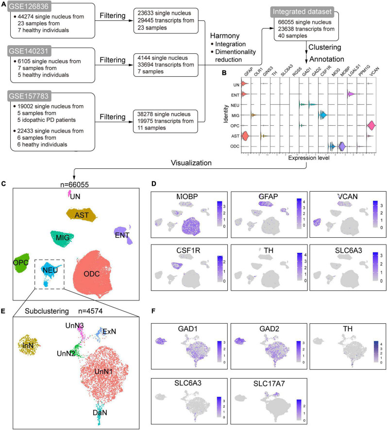
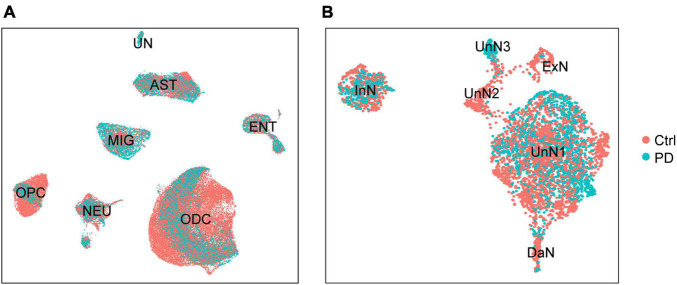
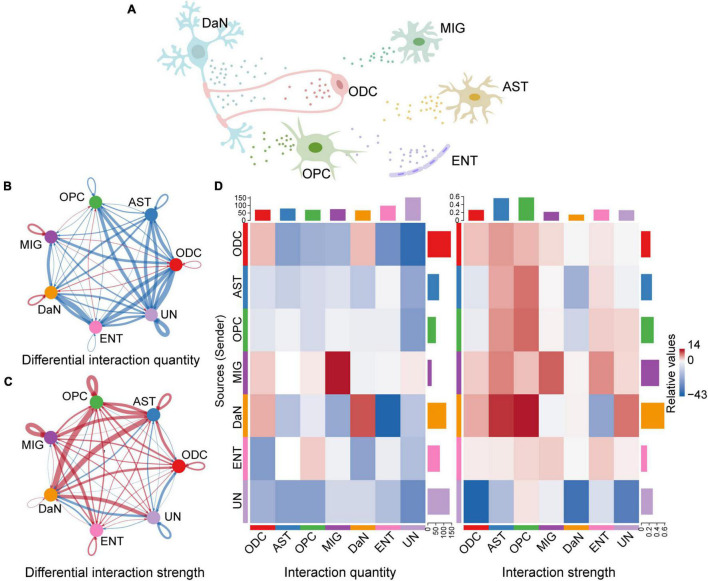
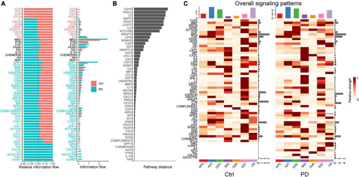
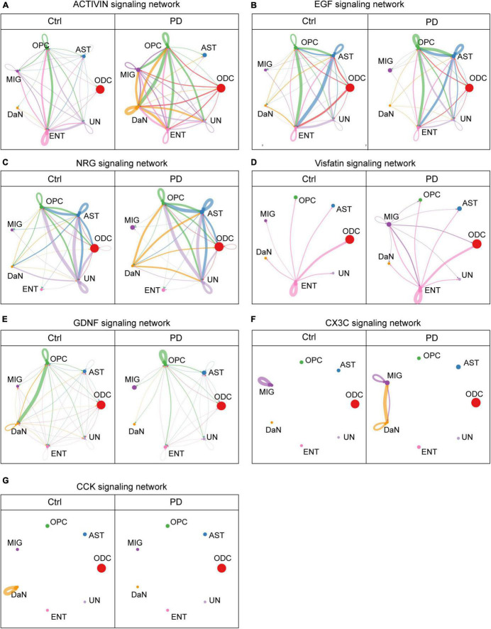
Parkinson's disease (PD) is a neurodegenerative movement disorder characterized with dopaminergic neuron (DaN) loss within the substantia nigra (SN). Despite bulk studies focusing on intracellular mechanisms of PD inside DaNs, few studies have explored the pathogeneses outside DaNs, or between DaNs and other cells. Here, we set out to probe the implication of intercellular communication involving DaNs in the pathogeneses of PD at a systemic level with bioinformatics methods. We harvested three online published single-cell/single-nucleus transcriptomic sequencing (sc/snRNA-seq) datasets of human SN (GSE126838, GSE140231, and GSE157783) from the Gene Expression Omnibus (GEO) database, and integrated them with one of the latest integration algorithms called Harmony. We then applied CellChat, the latest cell-cell communication analytic algorithm, to our integrated dataset. We first found that the overall communication quantity was decreased while the overall communication strength was enhanced in PD sample compared with control sample. We then focused on the intercellular communication where DaNs are involved, and found that the communications between DaNs and other cell types via certain signaling pathways were selectively altered in PD, including some growth factors, neurotrophic factors, chemokines, etc. pathways. Our bioinformatics analysis showed that the alteration in intercellular communications involving DaNs might be a previously underestimated aspect of PD pathogeneses with novel translational potential.
Keywords: CellChat; Parkinson’s disease; cell–cell communication; dopaminergic neurons; sc/snRNA-seq; signaling.
Copyright © 2022 Huang, Xu, Liu, Huang, Tan and Chen.
Conflict of interest statement
The authors declare that the research was conducted in the absence of any commercial or financial relationships that could be construed as a potential conflict of interest.
Figures
Similar articles
-
Single-Cell Nuclear Sequencing Atlas Revealed the Induction of Parkinson's Disease by RELN+ Neuron 3 and the Gene Regulatory Network of MSRA.Curr Med Chem. 2024 Mar 29. doi: 10.2174/0109298673289286240322041842. Online ahead of print. Curr Med Chem. 2024. PMID: 38561620
-
Analysis of gene expression in Parkinson's disease: possible involvement of neurotrophic support and axon guidance in dopaminergic cell death.Brain Pathol. 2009 Jan;19(1):91-107. doi: 10.1111/j.1750-3639.2008.00171.x. Epub 2008 May 7. Brain Pathol. 2009. PMID: 18462474 Free PMC article.
-
Construction of Parkinson's disease marker-based weighted protein-protein interaction network for prioritization of co-expressed genes.Gene. 2019 May 20;697:67-77. doi: 10.1016/j.gene.2019.02.026. Epub 2019 Feb 16. Gene. 2019. PMID: 30776463
-
Single-Cell RNA Sequencing in Parkinson's Disease.Biomedicines. 2021 Apr 1;9(4):368. doi: 10.3390/biomedicines9040368. Biomedicines. 2021. PMID: 33916045 Free PMC article. Review.
-
Integration of functional genomics data to uncover cell type-specific pathways affected in Parkinson's disease.Biochem Soc Trans. 2021 Nov 1;49(5):2091-2100. doi: 10.1042/BST20210128. Biochem Soc Trans. 2021. PMID: 34581766 Free PMC article. Review.
Cited by
-
Association of Serum Extracellular Vesicle miRNAs with Cognitive Functioning and Quality of Life in Parkinson's Disease.Biomolecules. 2024 Aug 13;14(8):1000. doi: 10.3390/biom14081000. Biomolecules. 2024. PMID: 39199388 Free PMC article.
-
Integrated analysis reveals the dysfunction of intercellular communication and metabolic signals in dilated cardiomyopathy.Heliyon. 2024 Feb 22;10(5):e26803. doi: 10.1016/j.heliyon.2024.e26803. eCollection 2024 Mar 15. Heliyon. 2024. PMID: 38434389 Free PMC article.
-
A Transcriptomic Roadmap of Parkinson's Disease Progression at Single Cell Resolution.medRxiv [Preprint]. 2025 Jul 30:2025.07.30.25332436. doi: 10.1101/2025.07.30.25332436. medRxiv. 2025. PMID: 40766144 Free PMC article. Preprint.
-
A Review of Single-Cell RNA-Seq Annotation, Integration, and Cell-Cell Communication.Cells. 2023 Jul 30;12(15):1970. doi: 10.3390/cells12151970. Cells. 2023. PMID: 37566049 Free PMC article. Review.
-
Identification of biomarkers in Parkinson's disease by comparative transcriptome analysis and WGCNA highlights the role of oligodendrocyte precursor cells.Front Aging Neurosci. 2024 Nov 20;16:1485722. doi: 10.3389/fnagi.2024.1485722. eCollection 2024. Front Aging Neurosci. 2024. PMID: 39634657 Free PMC article.
References
LinkOut - more resources
Full Text Sources
Miscellaneous