Review
. 2022 Feb 25:14:778404.
doi: 10.3389/fnagi.2022.778404. eCollection 2022.

Variability in Retinal Neuron Populations and Associated Variations in Mass Transport Systems of the Retina in Health and Aging

Affiliations
Review

Variability in Retinal Neuron Populations and Associated Variations in Mass Transport Systems of the Retina in Health and Aging

Moussa A Zouache. Front Aging Neurosci. .

Abstract

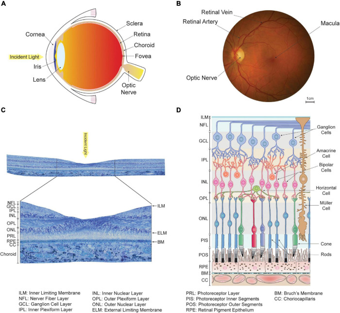
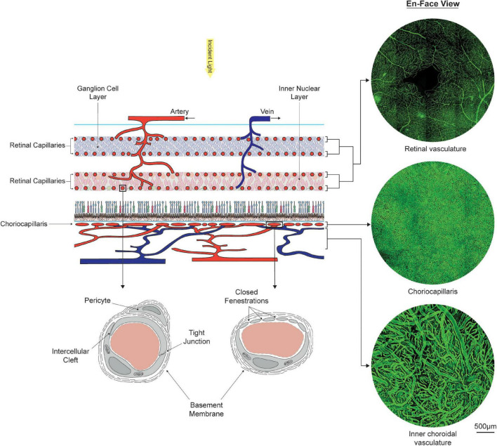
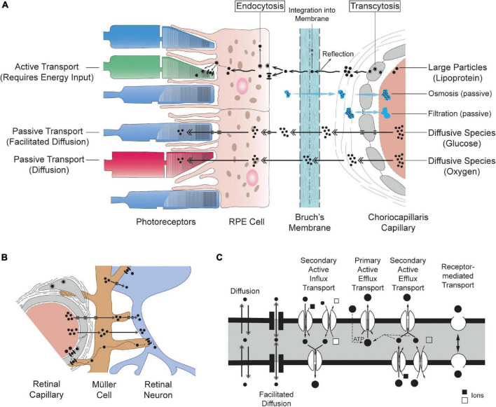
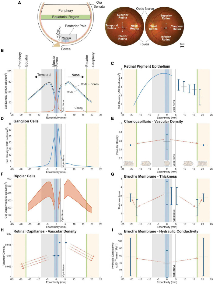
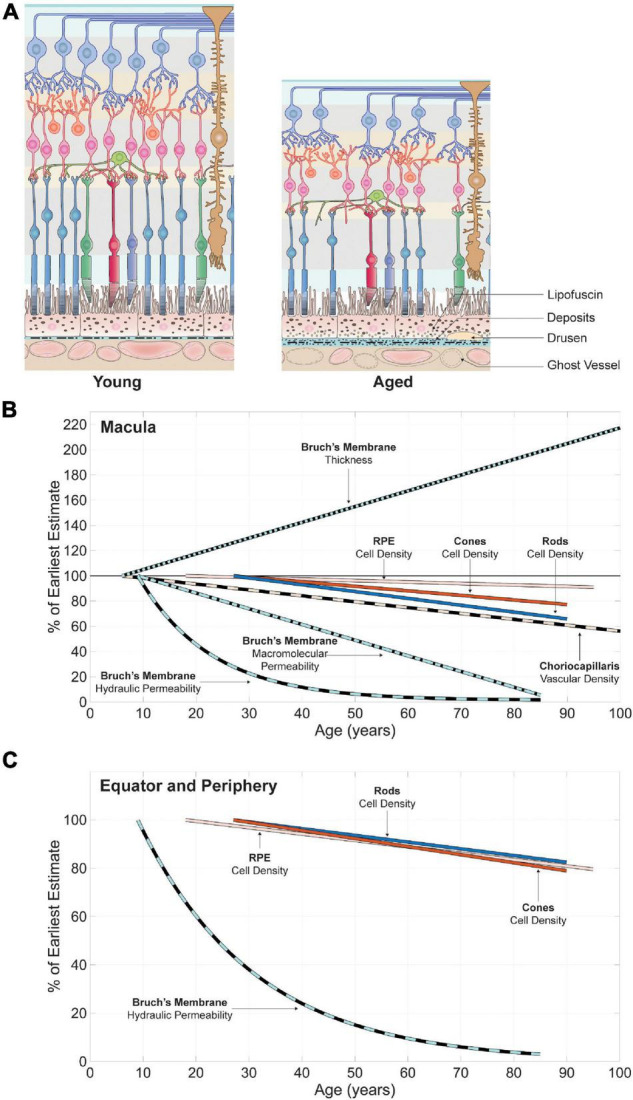
Aging is associated with a broad range of visual impairments that can have dramatic consequences on the quality of life of those impacted. These changes are driven by a complex series of alterations affecting interactions between multiple cellular and extracellular elements. The resilience of many of these interactions may be key to minimal loss of visual function in aging; yet many of them remain poorly understood. In this review, we focus on the relation between retinal neurons and their respective mass transport systems. These metabolite delivery systems include the retinal vasculature, which lies within the inner portion of the retina, and the choroidal vasculature located externally to the retinal tissue. A framework for investigation is proposed and applied to identify the structures and processes determining retinal mass transport at the cellular and tissue levels. Spatial variability in the structure of the retina and changes observed in aging are then harnessed to explore the relation between variations in neuron populations and those seen among retinal metabolite delivery systems. Existing data demonstrate that the relation between inner retinal neurons and their mass transport systems is different in nature from that observed between the outer retina and choroid. The most prominent structural changes observed across the eye and in aging are seen in Bruch's membrane, which forms a selective barrier to mass transfers at the interface between the choroidal vasculature and the outer retina.

Keywords: Bruch’s membrane; aging; choriocapillaris; mass transport; photoreceptors; retina; retinal neurons; retinal vasculature.


Conflict of interest statement

The author declares that the research was conducted in the absence of any commercial or financial relationships that could be construed as a potential conflict of interest.

Figures

FIGURE 1
FIGURE 1
Organization of the human eye and retina. (A) Schematic of a human eye. The anterior segments, which include the cornea, iris and lens, direct light toward the retina located in the back of the eye. (B) En-face view of the retina captured in a healthy individual using an ophthalmoscope. The optic nerve and large retinal arteries and veins are visible, but the underlying choroidal vasculature is not. The fovea, a region of the retina specialized for high-acuity and color vision, lies at the center of the approximately 5.5 mm wide macula. (C) View of a transverse section of the retina taken from a human donor eye in the macula. Histologically, the retina appears as layered tissue formed by retinal neurons, endothelium, and glial cells. (D) Schematic of the cellular organization of the retina and choriocapillaris, adapted and modified with permission from Campello et al. (2021). The location of histologically defined retinal layers is indicated. The outer retina consists of the retinal pigment epithelium and photoreceptor outer and inner segments. The inner retina includes horizontal, bipolar, amacrine and ganglion cells, which are all involved in the processing of signals originating from the photoreceptors.
FIGURE 2
FIGURE 2
Organization, morphology, and ultrastructure of the dual circulatory systems of the retina formed by the retinal and choroidal vasculatures. The inner two-thirds of the retina is sustained by at least four layers of retinal capillaries, which connect alternating arterial and venous branches. Oxygen and nutrients are supplied to the outer one third of the retina by the choriocapillaris, located externally to the retinal pigment epithelium. En-face visualizations of the retinal and choroidal vasculatures at the fovea were obtained by immunostaining portions of retinas and choroid with Ulex Europaeus Agglutinin and imaging them using confocal microscopy. Stark differences in vascular density between the choriocapillaris and the retinal capillary bed can be observed. Ultrastructural differences are also present. Retinal capillaries form a tightly regulated barrier between blood and tissue. Capillaries from the choriocapillaris present with closed fenestrations, which facilitate the transfer of small and large molecules.
FIGURE 3
FIGURE 3
Schematic representation of mass transport systems of the retina. The movement of molecules shown in (A,B) is directed from vasculatures to tissue only for simplicity; although, displayed mechanisms are also applicable to the opposite direction. (A) Simplified schematic of the transfer of material across choriocapillaris endothelial cells, Bruch’s membrane, RPE cells and photoreceptors (not to scale). (B) Mass transfers in the inner retina illustrating the symbiotic relationship between retinal endothelial cells, glial cells (here a Müller cell) and retinal neurons. (C) Schematic of the mechanisms of transport across cells adapted and modified from Hosoya and Tachikawa (2012). Transport is traditionally classified as passive (diffusion), carrier-mediated (facilitated diffusion, primary active efflux and secondary active influx and efflux) or receptor-mediated. The conventions used to depict mechanisms of transport in (A,B) are consistent with (C).
FIGURE 4
FIGURE 4
Spatial variations in retinal neuron populations (B,D,F) and in the key structures of their respective metabolite delivery systems (C,E,G–I). The approximate location of sampled retinal regions and their associated denomination is described in (A). Terminologies vary between studies; the ones employed here are consistent with the main text. Plotted data were collected from publications listed in Supplementary Table 1. All spatial variations are displayed along the nasal-temporal axis as illustrated in (B). Reported values are plotted as vertical line segments (ranges) or single points, and the approximate region they apply to is delineated using horizontal dashed lines. Dashed arrows indicate qualitatively reported trends. The density of bipolar cells (F) is plotted as a range inferred from approximate ratios between their density and that of their respective photoreceptor type. The maximal vascular density of retinal capillaries (H) appears to overlap with the highest density of bipolar (F) and ganglion (D) cells. Bruch’s membrane is thickest at the fovea (G), where photoreceptor density (B) and choriocapillaris vascular density (E) are highest.
FIGURE 5
FIGURE 5
Age-related variations in retinal neuron populations and in the key structures of their respective metabolite delivery systems in the macula and equator and periphery. A schematic of changes affecting the retina and choroid adapted and modified with permission from Campello et al. (2021) is shown in (A). Linear and non-linear regressions were collected from publications listed in Supplementary Table 2. The features plotted are normalized by the first value of each regression, which corresponds to the estimate in the youngest eye included in each study. In the macula (B), there can be large inter-individual differences that mask variations in the density of RPE cells and cone photoreceptors with age. With careful elimination of disease and other extraneous variables, some older subjects have significantly lower cone densities than do younger subjects. Aging is associated with an increase in the thickness of Bruch’s membrane and a reduction in its hydraulic and macromolecular permeability. The rate of rod loss and the decrease in the vascular density of the choriocapillaris observed with age lie within a similar range. In the equatorial and peripheral region (C) loss of cones with age is more pronounced as compared to rods.

Similar articles

Cited by

References

    1. Aalkjær C., Boedtkjer D., Matchkov V. (2011). Vasomotion - what is currently thought? Acta Physiol. 202 253–269. 10.1111/j.1748-1716.2011.02320.x - DOI - PubMed
    1. Abukawa H., Tomi M., Kiyokawa J., Hori S., Kondo T., Terasaki T., et al. (2009). Modulation of retinal capillary endothelial cells by Müller glial cell-derived factors. Mol. Vis. 15 451–457. - PMC - PubMed
    1. Aggarwal P., Nag T. C., Wadhwa S. (2007). Age-related decrease in rod bipolar cell density of the human retina: an immunohistochemical study. J. Biosci. 32 293–298. 10.1007/s12038-007-0029-9 - DOI - PubMed
    1. Ahnelt P. K., Kolb H., Pflug R. (1987). Identification of a subtype of cone photoreceptor, likely to be blue sensitive, in the human retina. J. Comp. Neurol. 255 18–34. 10.1002/cne.902550103 - DOI - PubMed
    1. Alberts B., Johnson A., Lewis J., Raff M., Roberts K., Walter P. (2014). Molecular biology of the cell, 6th Edn. New York, NY: Garland Science, 10.1201/9780203833445 - DOI

LinkOut - more resources