Review
. 2022 Feb 24:13:826314.
doi: 10.3389/fphys.2022.826314. eCollection 2022.

The Molecular Brakes of Adipose Tissue Lipolysis

Affiliations
Review

The Molecular Brakes of Adipose Tissue Lipolysis

Yongguo Li et al. Front Physiol. .

Abstract

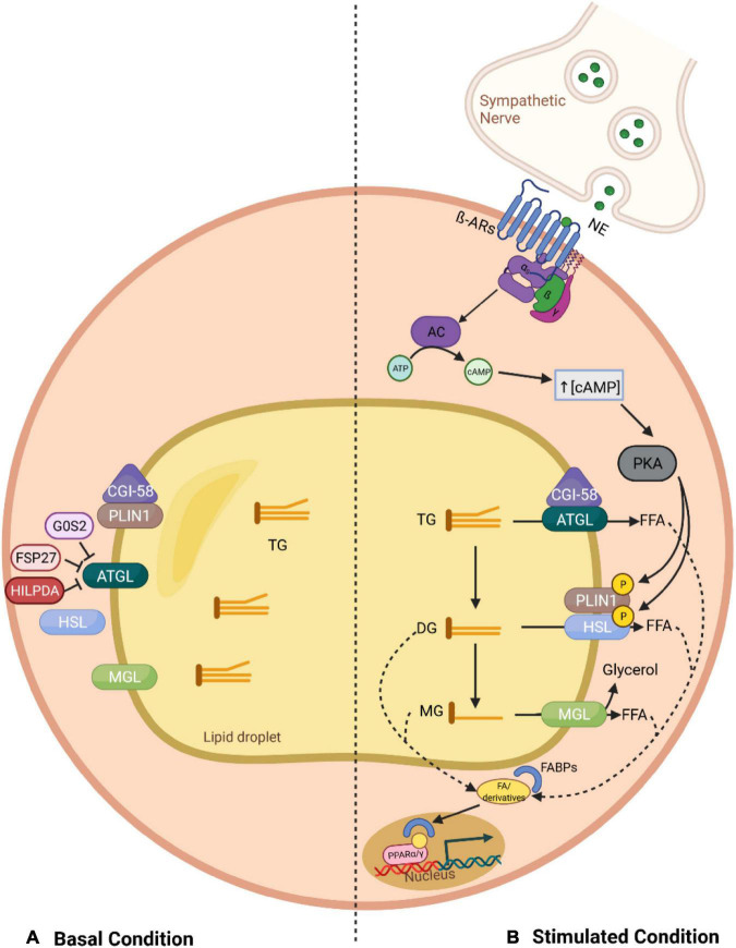
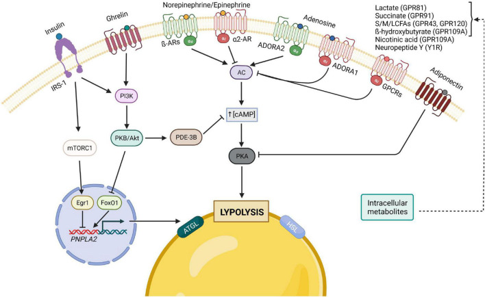
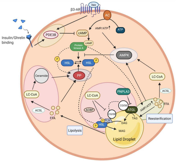
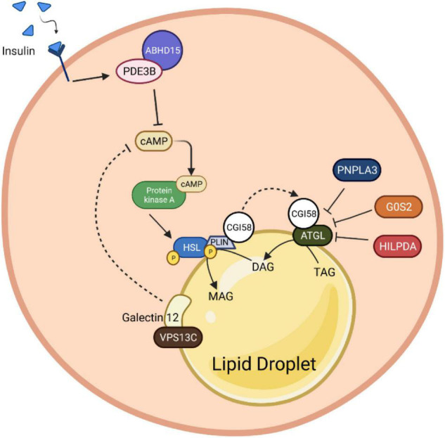
Adaptation to changes in energy availability is pivotal for the survival of animals. Adipose tissue, the body's largest reservoir of energy and a major source of metabolic fuel, exerts a buffering function for fluctuations in nutrient availability. This functional plasticity ranges from energy storage in the form of triglycerides during periods of excess energy intake to energy mobilization via lipolysis in the form of free fatty acids for other organs during states of energy demands. The subtle balance between energy storage and mobilization is important for whole-body energy homeostasis; its disruption has been implicated as contributing to the development of insulin resistance, type 2 diabetes and cancer cachexia. As a result, adipocyte lipolysis is tightly regulated by complex regulatory mechanisms involving lipases and hormonal and biochemical signals that have opposing effects. In thermogenic brown and brite adipocytes, lipolysis stimulation is the canonical way for the activation of non-shivering thermogenesis. Lipolysis proceeds in an orderly and delicately regulated manner, with stimulation through cell-surface receptors via neurotransmitters, hormones, and autocrine/paracrine factors that activate various intracellular signal transduction pathways and increase kinase activity. The subsequent phosphorylation of perilipins, lipases, and cofactors initiates the translocation of key lipases from the cytoplasm to lipid droplets and enables protein-protein interactions to assemble the lipolytic machinery on the scaffolding perilipins at the surface of lipid droplets. Although activation of lipolysis has been well studied, the feedback fine-tuning is less well appreciated. This review focuses on the molecular brakes of lipolysis and discusses some of the divergent fine-tuning strategies in the negative feedback regulation of lipolysis, including delicate negative feedback loops, intermediary lipid metabolites-mediated allosteric regulation and dynamic protein-protein interactions. As aberrant adipocyte lipolysis is involved in various metabolic diseases and releasing the brakes on lipolysis in thermogenic adipocytes may activate thermogenesis, targeting adipocyte lipolysis is thus of therapeutic interest.

Keywords: adipocytes; feedback mechanisms; free fatty acid; lipolysis; lipophagy; molecular brakes; reesterification; thermogenesis.


Conflict of interest statement

The authors declare that the research was conducted in the absence of any commercial or financial relationships that could be construed as a potential conflict of interest.

Figures

FIGURE 1
FIGURE 1
Overview of the canonical lipolysis pathway in adipocytes. (A) In the basal status, PLIN1 sequesters CGI-58, the co-activator of ATGL, limiting its function. This, together with the fact that ATGL can be further inhibited through direct interaction with G0S2 and FSP27, leads to a low TG hydrolysis rate. (B) In stimulated lipolysis, norepinephrine, secreted by sympathetic nerves innervating the adipose tissue, binds to β adrenergic receptors and activates AC. Consequently, there is an increase in the cytosolic levels of cAMP, which stimulates PKA, a kinase that phosphorylates PLIN1 and HSL. The phosphorylation of PLIN1 results in the release of CGI-58, which can then bind to ATGL, promoting the hydrolysis of TGs into DGs. In addition, upon phosphorylation of HSL, this lipase relocates to the lipid droplet surface where it can interact with PLIN1 and degrades DGs into MGs, which are further processed by MGL. Finally, the fatty acids generated in each lipolytic step, and their derivatives, can act as signaling ligands or as precursors for ligands of PPARα/γ and stimulate the expression of genes involved in energy homeostasis. AC, adenylyl cyclase; AR, adrenergic receptor; ATGL, adipose triglyceride lipase; ATP, adenosine triphosphate; cAMP, cyclic adenosine monophosphate; CGI-58, comparative gene identification 58; DG, diglyceride; FFA, free fatty acid; FABP, fatty acid-binding protein; FSP27, fat specific protein 27; G0S2, G0/S2 switch protein 2; HILPDA, hypoxia-induced lipid droplet-associated protein; HSL, hormone-sensitive lipase; MG, monoglyceride; MGL, monoglyceride lipase; NE, norepinephrine; PKA, protein kinase A; PLIN1, perilipin 1; PPAR, peroxisome proliferator-activated receptor; TG, triglyceride.
FIGURE 2
FIGURE 2
Principal hormonal, autocrine/paracrine modulators of adipocyte lipolysis. The binding of insulin to its receptor, IRS-1, leads to the activation of both PI3K/Akt and mTORC1 pathways, which result in the inhibition of lipolysis, either through the decrease of cAMP levels by PDE-3B (PI3K pathway) or by inhibiting the transcription of ATGL. The gut hormone ghrelin is also considered anti-lipolytic since it stimulates the PI3K pathway as well. Furthermore, lipolysis can also be inhibited by adiponectin, which inhibits PKA, through the reduction of its regulatory subunit of RIIα. In contrast, many of lipolysis’ regulators act on the pro-lipolytic PKA pathway, which is typically activated by catecholamines and can be inhibited by different factors upon binding to their respective Gi-coupled receptors. Of note, catecholamines and adenosine can both have pro- and anti-lipolytic effects depending on which receptor is predominantly expressed. AC, adenylyl cyclase; AR, adrenergic receptor; ATGL, adipose triglyceride lipase; cAMP, cyclic adenosine monophosphate; Egr1, Early growth response protein 1; FoxO1, forkhead box protein O1; GPCRs, G-protein-coupled receptors; HSL, hormone-sensitive lipase; IRS, insulin receptor substrate; mTORC1, mammalian target of rapamycin complex 1; PDE-3B, phosphodiesterase 3B; PI3K, phosphoinositide 3-kinase; PKA, protein kinase A; PKB, protein kinase B; NE/E, norepinephrine/epinephrine; ADORA1, adenosine receptor A1; ADORA2, adenosine receptor A2; S/M/LCFA, short-/medium-/long-chain fatty acid.
FIGURE 3
FIGURE 3
Intracellular networks regulate adipocyte lipolysis brake. The figure illustrates five feedback mechanism loops of lipolysis marked with different colors. Insulin and isoproterenol regulate lipolysis inhibition through PDE3B-mediated cAMP degradation (yellow circle). The fine-tuning of cAMP activation involves AMPK to induce a negative feedback loop in lipolysis. AMPK stimulation is coordinated via interaction with LC-CoA, which results from ACSL-mediated FFA conversion (light blue circle). Besides LC-CoA, cAMP production and degradation together with re-esterification, participate in this AMPK induction due to an increase of the AMP/ATP ratio yielded during these processes. PKA can potentially activate a re-eseterification enzyme called DGAT (green circle). Elevated AMPK promotes a negative feedback loop of lipolysis by inhibiting PKA-mediated HSL phosphorylation. In contrast to PKA, HSL phosphorylation can be reversed by PP, whose activation highly relies on the presence of FFA and PKA to induce a feedback mechanism (purple circle). The intermediary lipid metabolite, LC-CoA, can enter the ceramide biosynthesis pathway, followed by lipolysis attenuation via PP2A activation. The dynamic regulation of endogenous LC-CoA is also shown through its impact on protein lipases (HSL, ATGL) depletion. LC-CoA acts as a non-competitive inhibitor, where it binds to CGI58 and interacts with PLIN. The LC-CoA-CGI58-PLIN complex results in the deactivation of ATGL. LC-CoA also impairs HSL function enhanced by ACBP. Similar to LC-CoA, PNPLA3 serves as a competitive inhibitor of CGI58 to attenuate the ATGL catalytic activity (pink circle). PDE3B, phosphodiesterase 3B; cAMP, cyclic adenosine monophosphate; PKA, protein kinase A; AMPK, AMP-activated protein kinase; DGAT, diglyceride acyltransferase; LC-CoA, long-chain fatty acyl–CoA esters; ACSL, long-chain-fatty-acid—CoA ligase; FFA, free fatty acid; AMP/ATP, adenosine monophosphate/adenosine triphosphate ratio; HSL, hormone-sensitive lipase; PP2A, protein phosphatase 2A; ATGL, adipose triglyceride lipase; CGI-58, comparative gene identification 58; PLIN1, perilipin 1; ACBP, acyl-CoA-binding protein; PNPLA3, patatin-like phospholipase domain-containing protein 3.
FIGURE 4
FIGURE 4
Protein-protein interaction roles in adipocyte lipolysis. Insulin binding induces stabilization of PDE3B with ABHD15. This molecular complex promotes cAMP degradation, which consequently inhibits the HSL phosphorylation upon PKA inactivation. Besides HSL, PLIN1 is also directly regulated by the PKA. The stimulation of PLIN1 mediates CGI58 release from the PLIN1 and HSL complex to bind with ATGL and induce its catalytic activity. Nevertheless, the complex of CGI58 and HSL can be suppressed by the presence of binding inhibitors, G0S2, HILPDA and PNPLA3. Presumably, Galectin12, a protein that localizes on lipid droplets, and its coactivator, VPSC13, can alter the cAMP production by inhibiting the PDE recruitment function, which in the end contributes to lipolysis abolition. PDE3B, phosphodiesterase 3B; cAMP, cyclic adenosine monophosphate; AMPK, AMP-activated protein kinase; HSL, hormone-sensitive lipase; PLIN1, perilipin 1; PKA, protein kinase A; CGI-58, comparative gene identification 58; ATGL, adipose triglyceride lipase; G0S2, G0/S2 switch protein 2; HILPDA, hypoxia-induced lipid droplet-associated protein; PNPLA3, patatin-like phospholipase domain-containing protein 3; VPSC13, vacuolar protein sorting 13C.
FIGURE 5
FIGURE 5
A schematic diagram of fine-tuned lipolysis in thermogenic biology. Upon cold stimulation, the non-shivering thermogenesis process in BAT can be mediated through intra- and extra-BAT FFAs source. In local lipolysis, FFAs generated from brown adipocyte lipid droplets can be used directly to activate and fuel UCP1 to produce heat. In addition, there is also a synergistic and compensatory mechanism, where white adipocyte lipolysis and TRL degradation (with the help of LPL and LAL) serve as extracellular fuels to permit a maximum thermogenesis level. These circulating FFAs are imported into BAT via FABP, FATP1 and CD36 transporters. Among them, FABP assists the binding of FFA released in WAT, which is later imported into brown adipocytes as a FABP-FFA complex via endocytosis. AR, adrenergic receptor; FFA, free fatty acid; NE, norepinephrine; FABP, fatty acid-binding protein; FATP, fatty acid transport proteins; TRL, triglyceride-rich lipoprotein; LPL, lipoprotein lipase; LAL, lysosomal acid lipase; UCP1, uncoupling protein 1.

References

    1. Agustsson T., Rydén M., Hoffstedt J., van Harmelen V., Dicker A., Laurencikiene J., et al. (2007). Mechanism of increased lipolysis in cancer cachexia. Cancer Res. 67 5531–5537. 10.1158/0008-5472.CAN-06-4585 - DOI - PubMed
    1. Ahmad F., Lindh R., Tang Y., Weston M., Degerman E., Manganiello V. C. (2007). Insulin-induced formation of macromolecular complexes involved in activation of cyclic nucleotide phosphodiesterase 3B (PDE3B) and its interaction with PKB. Biochem. J. 404 257–268. 10.1042/BJ20060960 - DOI - PMC - PubMed
    1. Ahmadian M., Abbott M. J., Tang T., Hudak C. S., Kim Y., Bruss M., et al. (2011). Desnutrin/ATGL is regulated by AMPK and is required for a brown adipose phenotype. Cell Metab. 13 739–748. 10.1016/j.cmet.2011.05.002 - DOI - PMC - PubMed
    1. Ahmadian M., Duncan R. E., Varady K. A., Frasson D., Hellerstein M. K., Birkenfeld A. L., et al. (2009). Adipose overexpression of desnutrin promotes fatty acid use and attenuates diet-induced obesity. Diabetes 58 855–866. 10.2337/db08-1644 - DOI - PMC - PubMed
    1. Ahmed K., Tunaru S., Tang C., Muller M., Gille A., Sassmann A., et al. (2010). An autocrine lactate loop mediates insulin-dependent inhibition of lipolysis through GPR81. Cell Metab. 11 311–319. 10.1016/j.cmet.2010.02.012 - DOI - PubMed