. 2022 Feb 22:13:804469.
doi: 10.3389/fphys.2022.804469. eCollection 2022.

Hypoxia During the Consolidation Phase of Distraction Osteogenesis Promotes Bone Regeneration

Affiliations

Hypoxia During the Consolidation Phase of Distraction Osteogenesis Promotes Bone Regeneration

Yanshi Liu et al. Front Physiol. .

Abstract

Background: Hypoxia is the critical driving force for angiogenesis and can trigger the osteogenic-angiogenic coupling followed by the enhancement of bone regeneration. While lots of studies showed that hypoxia administration can accelerate bone formation during distraction osteogenesis (DO), the therapeutic timing for the osteogenic purpose was concentrated on the distraction phase. The outcomes of hypoxia administration in the consolidation phase stay uncertain. The purpose of this study was to determine the osteogenic effectiveness of hypoxia therapy during the consolidation phase, if any, to enhance bone regeneration in a rat femoral DO model.

Methods: A total of 42 adult male Sprague-Dawley rats underwent right femoral mid-diaphysis transverse osteotomy and were randomly divided into Control (NS administration, n = 21) and Group1 (deferoxamine therapy, n = 21) after distraction. During the consolidation phase, Group1 was treated with local deferoxamine (DFO) injection into the distraction zone, while the Control underwent the same dosage of NS. Animals were sacrificed after 2, 4, and 6 weeks of consolidation. The process of bone formation and remodeling was monitored by digital radiographs, and the regenerated bone was evaluated by micro-computed tomography (micro-CT), biomechanical test, and histological analysis. The serum content of hypoxia-inducible factor 1α (HIF-1α) and vascular endothelial growth factor (VEGF) were measured by enzyme linked immunosorbent assay (ELISA) for further analysis.

Results: Bone regeneration was significantly enhanced after hypoxia therapy during the consolidation phase. The digital radiograph, micro-CT, and biomechanical evaluation showed better effects regarding volume, continuity, and mechanical properties of the regenerated bone in Group1. The histomorphological evaluation also revealed the hypoxia treatment contributed to accelerate bone formation and remodeling during DO. The higher positive expression of angiogenic and osteogenic markers were observed in Group1 after hypoxia administration according to the immunohistochemical analysis. The serum content of HIF-1α and VEGF was also increased after hypoxia therapy as evidenced from ELISA.

Conclusion: Hypoxia administration during the consolidation phase of distraction osteogenesis has benefits in enhancing bone regeneration, including accelerates the bone formation and remodeling.

Keywords: bone regeneration; consolidation; distraction osteogenesis; hypoxia; osteogenic-angiogenic coupling.


Conflict of interest statement

The authors declare that the research was conducted in the absence of any commercial or financial relationships that could be construed as a potential conflict of interest.

Figures
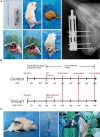
FIGURE 1
FIGURE 1
Methodologies of the present study. (A) The surgical procedures for the right femur model of distraction osteogenesis. (B) Rats were divided into two groups according to different interventions. DFO injection or NS administration as control was started from postoperative days 16 to 26. (C) Under image intensifier control, the solution was locally injected into the distraction gap every other day during the consolidation phase.
FIGURE 2
FIGURE 2
Hypoxia therapy during the consolidation phase accelerated bone regeneration in a rat femoral distraction osteogenesis model. The yellow arrows indicate bone union with primary recanalization of the medullary cavity was achieved in Group1 after 6 weeks of consolidation. (A) X-ray images of the distraction zone weekly until a 6-week consolidation duration was completed. (B) The general images of specimens after 2, 4, and 6 weeks of consolidation.
FIGURE 3
FIGURE 3
Results of micro-CT evaluation demonstrating promotion in regenerate quality after hypoxia therapy during the consolidation phase. (A) Representative 3D micro-CT images of the distraction zone at the termination of the 6-week consolidation. (B,C) Quantitative evaluation of BMD and BV/TV (n = 3 per group), manifesting both of the two values in Group1 were significantly higher than those in control (*p < 0.05, **P < 0.01, independent t-test).
FIGURE 4
FIGURE 4
Results of mechanical properties and values were normalized to the contralateral femur (n = 3 per group) (*p < 0.05, **P < 0.01, independent t-test).
FIGURE 5
FIGURE 5
Histological analysis of bone regeneration during the consolidation phase. Von Kossa, Masson Trichrome, Goldner Trichrome, and Safranin O staining indicated the enhanced bone regeneration in the hypoxia therapy group (Group1). (A) Representative images of the histology sections. (B) Quantification of the ratio of regenerated bone in the distraction zone (*p < 0.05, **P < 0.01, independent t-test).
FIGURE 6
FIGURE 6
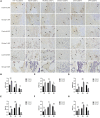
(A) Immunohistochemistry images of HIF-1α, VEGF, RUNX2, Osterix, OCN, and OPN in the two groups at the termination of 2-week, 4-week, and 6-week consolidation. Black arrows, positive cells in Control. Dotted arrows, positive cells in Group1. (B–G) The semiquantitative measurements (n = 3 per group) showed the 6 markers were highly expressed in Group1 compared to Control in the first 4 weeks consolidation duration. At the termination of the 6-week consolidation, HIF-1α and VEGF were expressed higher in Group1 than in Control, RUNX2 was expressed lower in Group1 than in Control, and the differences of the other three markers were not statistically significant (*p < 0.05, **P < 0.01, independent t-test).
FIGURE 7
FIGURE 7
Serum content of HIF-1α and VEGF evaluated by ELISA (n = 3 per group). During the consolidation phase, the serum content of HIF-1α and VEGF were higher in Group1 compared to Control after 2 weeks. After 4 weeks, VEGF content was higher in Group1 than that in Control, while HIF-1α content was similar between the two groups. There was no statistical significance in the level of HIF-1α and VEGF between the two groups after 6 weeks (*p < 0.05, **P < 0.01, independent t-test).

References

    1. Ai-Aql Z. S., Alagl A. S., Graves D. T., Gerstenfeld L. C., Einhorn T. A. (2008). Molecular mechanisms controlling bone formation during fracture healing and distraction osteogenesis. J. Dent Res. 87 107–118. 10.1177/154405910808700215 - DOI - PMC - PubMed
    1. Alzahrani M. M., Anam E., AlQahtani S. M., Makhdom A. M., Hamdy R. C. (2018). Strategies of enhancing bone regenerate formation in distraction osteogenesis. Connect Tissue Res. 59 1–11. - PubMed
    1. Arnett T. R. (2010). Acidosis, hypoxia and bone. Arch. Biochem. Biophys. 503 103–109. 10.1016/j.abb.2010.07.021 - DOI - PubMed
    1. Aronson J. (1994). Temporal and spatial increases in blood flow during distraction osteogenesis. Clin. Orthop. Relat. Res. 1994 124–131. 10.1097/00003086-199404000-00020 - DOI - PubMed
    1. Baek W. Y., Lee M. A., Jung J. W., Kim S. Y., Akiyama H., de Crombrugghe B., et al. (2009). Positive regulation of adult bone formation by osteoblast-specific transcription factor osterix. J. Bone Miner. Res. 24 1055–1065. 10.1359/jbmr.081248 - DOI - PMC - PubMed