. 2022 Feb 5;12(3):e4305.
doi: 10.21769/BioProtoc.4305.

Transient Middle Cerebral Artery Occlusion with an Intraluminal Suture Enables Reproducible Induction of Ischemic Stroke in Mice

Affiliations

Transient Middle Cerebral Artery Occlusion with an Intraluminal Suture Enables Reproducible Induction of Ischemic Stroke in Mice

Luke R Lemmerman et al. Bio Protoc. .

Abstract

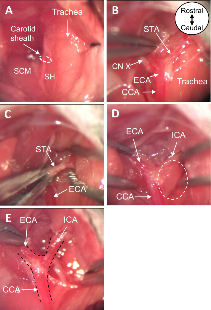
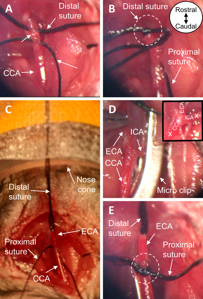
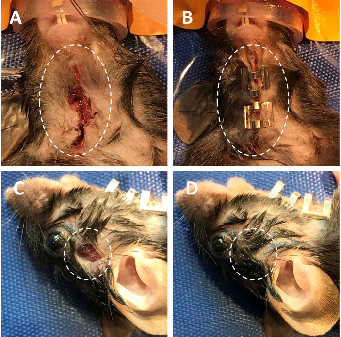
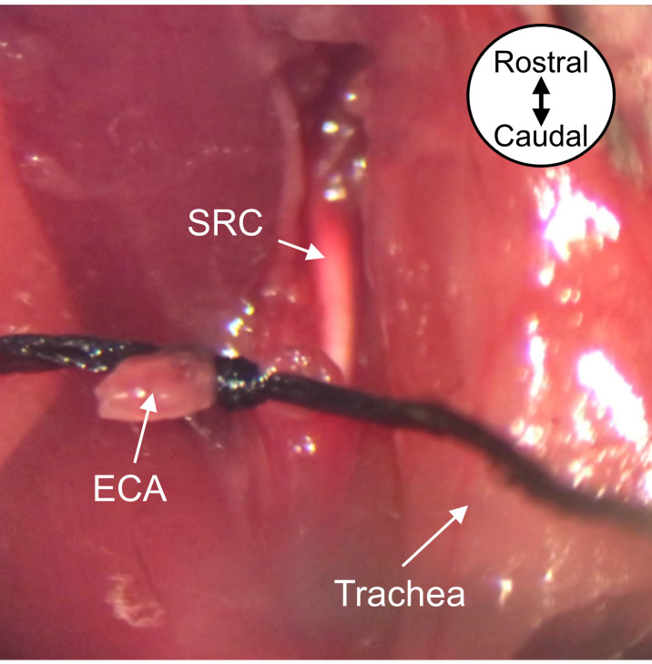
Ischemic stroke is a leading cause of mortality and chronic disability worldwide, underscoring the need for reliable and accurate animal models to study this disease's pathology, molecular mechanisms of injury, and treatment approaches. As most clinical strokes occur in regions supplied by the middle cerebral artery (MCA), several experimental models have been developed to simulate an MCA occlusion (MCAO), including transcranial MCAO, micro- or macro-sphere embolism, thromboembolisation, photothrombosis, Endothelin-1 injection, and - the most common method for ischemic stroke induction in murine models - intraluminal MCAO. In the intraluminal MCAO model, the external carotid artery (ECA) is permanently ligated, after which a partially-coated monofilament is inserted and advanced proximally to the common carotid artery (CCA) bifurcation, before being introduced into the internal carotid artery (ICA). The coated tip of the monofilament is then advanced to the origin of the MCA and secured for the duration of occlusion. With respect to other MCAO models, this model offers enhanced reproducibility regarding infarct volume and cognitive/functional deficits, and does not require a craniotomy. Here, we provide a detailed protocol for the surgical induction of unilateral transient ischemic stroke in mice, using the intraluminal MCAO model. Graphic abstract: Overview of the intraluminal monofilament method for transient middle cerebral artery occlusion (MCAO) in mouse.

Keywords: Brain reperfusion; Cerebral infarct; Intraluminal MCAO; Murine model; Rodent model; Transient MCAO.


Conflict of interest statement

Competing interestsNo potential competing interest was reported by the authors.

Figures

Figure 1.
Figure 1.. Make incision for LDF fiber.
A. Prepare area between the right eye and right ear for incision. B. make a small incision in the prepped area. LDF, Laser Doppler Flowmetry.
Figure 2.
Figure 2.. Make ventral neck incision and place LDF fiber against the skull.
A. Lateral and B. ventral views of the neck incision pulled open with braided sutures, with the LDF fiber placed against the skull. C. Zoomed view of ventral neck incision pulled open. LDF, Laser Doppler Flowmetry.
Figure 3.
Figure 3.. Expose CCA, ECA, and ICA.
A. View of ventral neck incision before and B. after blunt dissection. C. Cauterize the STA with bipolar forceps. D. Detach the superior cervical ganglion at the carotid body from the carotid arteries. E. View of exposed CCA, ECA, and ICA after dissection. All images are ventral views oriented from rostral to caudal as depicted in panel B. CCA, common carotid artery; ECA, external carotid artery; ICA, internal carotid artery; STA, superior thyroid artery; SCM, sternocleidomastoid muscle; SH, sternohyoid muscle; CN X, vagus nerve.
Figure 4.
Figure 4.. Prepare the carotid arteries for insertion of the monofilament.
A. Place two black braided silk sutures under the ECA. B. With one suture, permanently ligate the most distal end of the exposed ECA. C. Tape the ends of the suture to the nose cone, pulling the ECA taut. D. Place a micro clip across both the CCA and ICA, to block blood flow at the carotid bifurcation. E. Using the remaining suture, tie a loose knot around the proximal end of the ECA near its origin. All images are ventral views oriented from rostral to caudal as depicted in panel B. ECA, external carotid artery; CCA, common carotid artery; ICA, internal carotid artery.
Figure 5.
Figure 5.. Insert the monofilament into the ICA.
A. Make a small incision in the ECA and B. insert the coated tip of the monofilament. C. Tighten suture knot around ECA containing the coated tip. D. Remove the micro clip, restoring blood flow to the CCA and ICA, and E. transect the ECA at the monofilament entry site. F-G. Advance the monofilament by way of the CCA bifurcation into the ICA, carefully avoiding the PPA, until the monitored cortical blood flow decreases by 40-70% of baseline, H-I. typically correlating with an insertion length of 6.5-10 mm (distance delineated by gold marks in figure). All images are ventral views oriented from rostral to caudal as depicted in panel C. ECA, external carotid artery; CCA, common carotid artery; ICA, internal carotid artery; PPA, pterygopalatine artery; CN XII, hypoglossal nerve.
Figure 6.
Figure 6.. Close neck and scalp incisions.
A-B. Temporarily close the ventral neck incision using AutoClips (ventral view). C-D. Close scalp incision with tissue adhesive (lateral view).
Figure 7.
Figure 7.. Remove the monofilament to allow reperfusion of MCA-supplied tissue.
Remove the monofilament from the ECA and tighten the proximal ECA suture knot around the ECA stump. The silicone rubber coating from the monofilament tip will remain in the ICA. Image is a ventral view oriented from rostral to caudal as depicted. MCA, middle cerebral artery; ECA, external carotid artery; SRC, silicone rubber coating.
Video 1.
Video 1.. Surgical procedure for induction of ischemic stroke in a mouse model via intraluminal suture tMCAO.

References

    1. Adams H. P. Jr.and Nudo R. J.(2013). Management of patients with stroke: is it time to expand treatment options? Ann Neurol 74(1): 4-10. - PMC - PubMed
    1. Albers G. W., Marks M. P., Kemp S., Christensen S., Tsai J. P., Ortega-Gutierrez S., McTaggart R. A., Torbey M. T., Kim-Tenser M., Leslie-Mazwi T., et al. .(2018). Thrombectomy for Stroke at 6 to 16 Hours with Selection by Perfusion Imaging. N Engl J Med 378(8): 708-718. - PMC - PubMed
    1. Balch M. H. H., Harris H., Chugh D., Gnyawali S., Rink C., Nimjee S. M. and Arnold W. D.(2021). Ischemic stroke-induced polyaxonal innervation at the neuromuscular junction is attenuated by robot-assisted mechanical therapy. Exp Neurol 343: 113767. - PMC - PubMed
    1. Barber P. A., Hoyte L., Colbourne F. and Buchan A. M.(2004). Temperature-regulated model of focal ischemia in the mouse: a study with histopathological and behavioral outcomes. Stroke 35(7): 1720-1725. - PubMed
    1. Benjamin E. J., Muntner P., Alonso A., Bittencourt M. S., Callaway C. W., Carson A. P., Chamberlain A. M., Chang A. R., Cheng S., Das S. R., et al. .(2019). Heart Disease and Stroke Statistics-2019 Update: A Report From the American Heart Association. Circulation 139(10): e56-e528. - PubMed