

Cytopathic Effect Assay and Plaque Assay to Evaluate in vitro Activity of Antiviral Compounds Against Human Coronaviruses 229E, OC43, and NL63
- PMID: 35284599
- PMCID: PMC8855088
- DOI: 10.21769/BioProtoc.4314
Cytopathic Effect Assay and Plaque Assay to Evaluate in vitro Activity of Antiviral Compounds Against Human Coronaviruses 229E, OC43, and NL63
Abstract
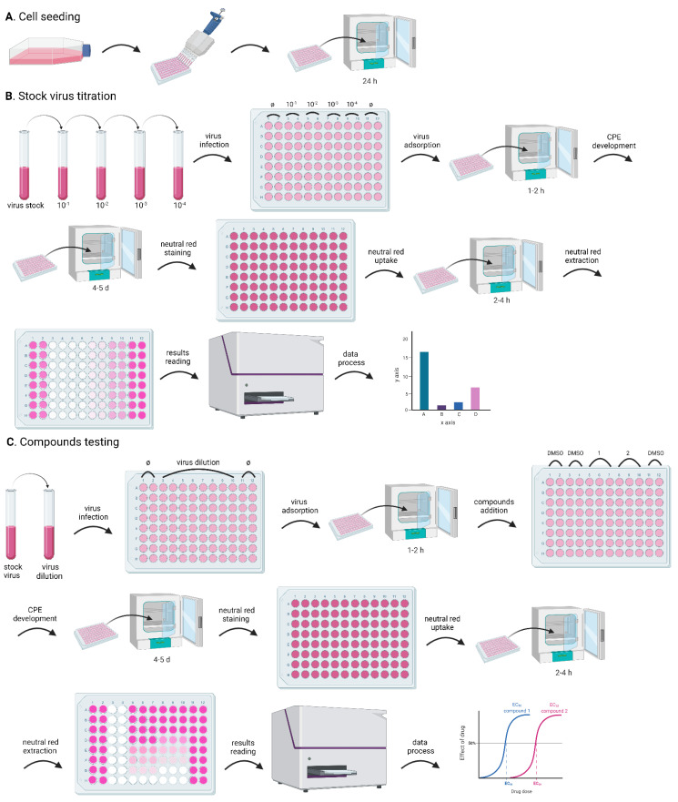
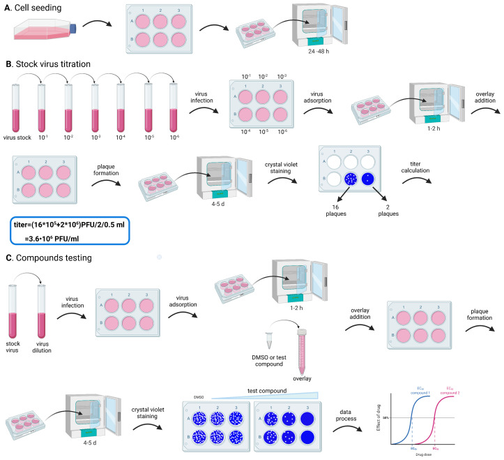
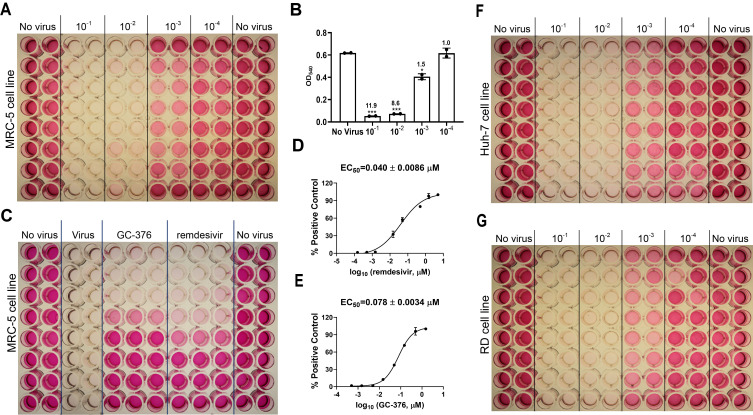
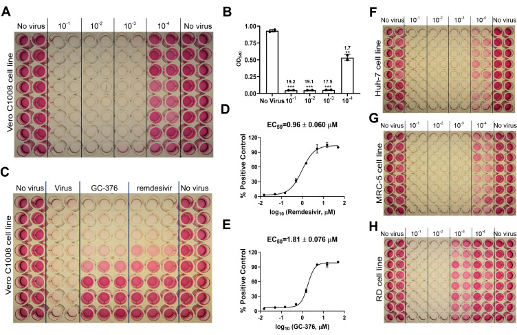
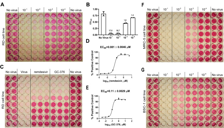
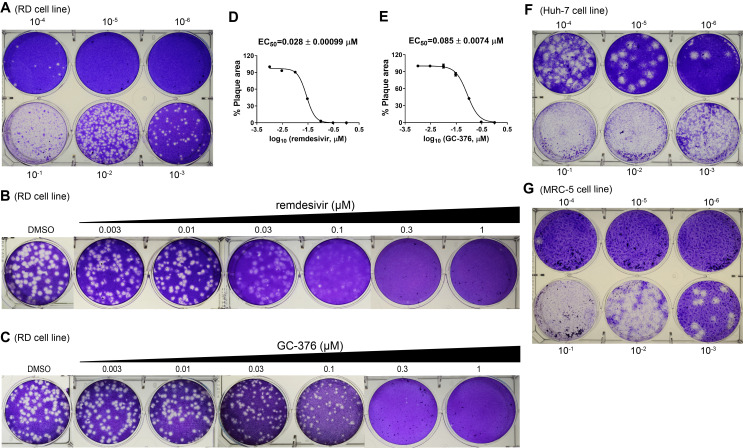
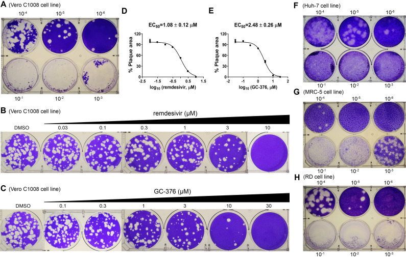
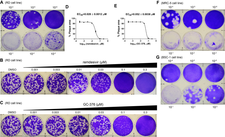
Coronaviruses are important human pathogens, among which the severe acute respiratory syndrome coronavirus 2 (SARS-CoV-2) is the causative agent for the COVID-19 pandemic. To combat the SARS-CoV-2 pandemic, there is a pressing need for antivirals, especially broad-spectrum antivirals that are active against all seven human coronaviruses (HCoVs). For this reason, we are interested in developing antiviral assays to expedite the drug discovery process. Here, we provide the detailed protocol for the cytopathic effect (CPE) assay and the plaque assay for human coronaviruses 229E (HCoV-229E), HCoV-OC43, and HCoV-NL63, to identify novel antivirals against HCoVs. Neutral red was used in the CPE assay, as it is relatively inexpensive and more sensitive than other reagents. Multiple parameters including multiplicity of infection, incubation time and temperature, and staining conditions have been optimized for CPE and plaque assays for HCoV-229E in MRC-5, Huh-7, and RD cell lines; HCoV-OC43 in RD, MRC-5, and BSC-1 cell lines, and HCoV-NL63 in Vero E6, Huh-7, MRC-5, and RD cell lines. Both CPE and plaque assays have been calibrated with the positive control compounds remdesivir and GC-376. Both CPE and plaque assays have high sensitivity, excellent reproducibility, and are cost-effective. The protocols described herein can be used as surrogate assays in the biosafety level 2 facility to identify entry inhibitors and protease inhibitors for SARS-CoV-2, as HCoV-NL63 also uses ACE2 as the receptor for cell entry, and the main proteases of HCoV-OC43 and SARS-CoV-2 are highly conserved. In addition, these assays can also be used as secondary assays to profile the broad-spectrum antiviral activity of existing SARS-CoV-2 drug candidates.
Keywords: 229E; Antiviral; Human Coronavirus; NL63; OC43.
Copyright © 2022 The Authors; exclusive licensee Bio-protocol LLC.
Conflict of interest statement
Competing interestsThe authors declare no competing interests.
Figures
Similar articles
-
Improved Culture Methods for Human Coronaviruses HCoV-OC43, HCoV-229E, and HCoV-NL63.Curr Protoc. 2023 Oct;3(10):e914. doi: 10.1002/cpz1.914. Curr Protoc. 2023. PMID: 37882768 Free PMC article.
-
Seasonal human coronaviruses OC43, 229E, and NL63 induce cell surface modulation of entry receptors and display host cell-specific viral replication kinetics.Microbiol Spectr. 2024 Jul 2;12(7):e0422023. doi: 10.1128/spectrum.04220-23. Epub 2024 Jun 12. Microbiol Spectr. 2024. PMID: 38864599 Free PMC article.
-
Improving human coronavirus OC43 (HCoV-OC43) research comparability in studies using HCoV-OC43 as a surrogate for SARS-CoV-2.J Virol Methods. 2022 Jan;299:114317. doi: 10.1016/j.jviromet.2021.114317. Epub 2021 Oct 9. J Virol Methods. 2022. PMID: 34634321 Free PMC article.
-
An overview on the seven pathogenic human coronaviruses.Rev Med Virol. 2022 Mar;32(2):e2282. doi: 10.1002/rmv.2282. Epub 2021 Aug 2. Rev Med Virol. 2022. PMID: 34339073 Review.
-
Current Strategies of Antiviral Drug Discovery for COVID-19.Front Mol Biosci. 2021 May 13;8:671263. doi: 10.3389/fmolb.2021.671263. eCollection 2021. Front Mol Biosci. 2021. PMID: 34055887 Free PMC article. Review.
Cited by
-
Allo-Priming Reverses Immunosenescence and May Restore Broad Respiratory Viral Protection and Vaccine Responsiveness to the Elderly: Results of a Phase I/II Clinical Trial.Vaccines (Basel). 2025 Apr 25;13(5):463. doi: 10.3390/vaccines13050463. Vaccines (Basel). 2025. PMID: 40432075 Free PMC article.
-
Safety and immunogenicity of SARS-CoV-2 protein subunit recombinant vaccine (Indovac®) in healthy populations aged 18 years and above in Indonesia: A phase I, observer-blind, randomized, controlled study.Hum Vaccin Immunother. 2025 Dec;21(1):2501467. doi: 10.1080/21645515.2025.2501467. Epub 2025 May 17. Hum Vaccin Immunother. 2025. PMID: 40381203 Free PMC article. Clinical Trial.
-
Comparative study of the propagation and plaque titration conditions for human coronavirus OC43 as a surrogate for SARS-CoV-2.Arch Virol. 2024 Oct 4;169(10):214. doi: 10.1007/s00705-024-06146-9. Arch Virol. 2024. PMID: 39365483
-
Utility of an In-Vitro Micro-Neutralizing Test in Comparison to a Plaque Reduction Neutralization Test for Dengue Virus, Japanese Encephalitis Virus, and Zika Virus Serology and Drug Screening.Pathogens. 2023 Dec 20;13(1):8. doi: 10.3390/pathogens13010008. Pathogens. 2023. PMID: 38276154 Free PMC article.
-
Optimizing human coronavirus OC43 growth and titration.PeerJ. 2022 Jul 8;10:e13721. doi: 10.7717/peerj.13721. eCollection 2022. PeerJ. 2022. PMID: 35833016 Free PMC article.
References
-
- Brussaard C. P., Marie D. and Bratbak G.(2000). Flow cytometric detection of viruses. J Virol Methods 85(1-2): 175-182. - PubMed
Grants and funding
LinkOut - more resources
Full Text Sources
Research Materials
Miscellaneous
