Review
. 2022 Mar 13;79(3):187.
doi: 10.1007/s00018-022-04220-6.

Hematopoietic responses to SARS-CoV-2 infection

Affiliations
Review

Hematopoietic responses to SARS-CoV-2 infection

Shokrollah Elahi. Cell Mol Life Sci. .

Abstract

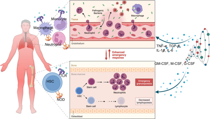
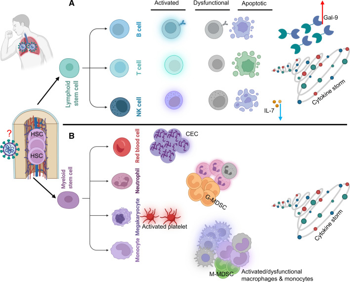
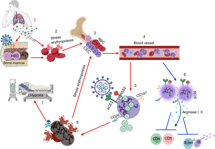
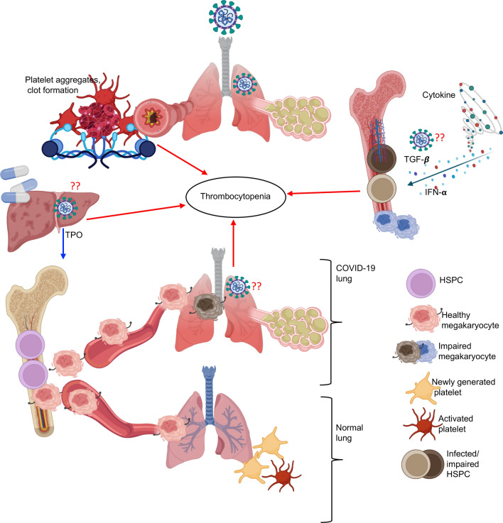
Under physiological conditions, hematopoietic stem and progenitor cells (HSPCs) in the bone marrow niches are responsible for the highly regulated and interconnected hematopoiesis process. At the same time, they must recognize potential threats and respond promptly to protect the host. A wide spectrum of microbial agents/products and the consequences of infection-induced mediators (e.g. cytokines, chemokines, and growth factors) can have prominent impact on HSPCs. While COVID-19 starts as a respiratory tract infection, it is considered a systemic disease which profoundly alters the hematopoietic system. Lymphopenia, neutrophilia, thrombocytopenia, and stress erythropoiesis are the hallmark of SARS-CoV-2 infection. Moreover, thrombocytopenia and blood hypercoagulability are common among COVID-19 patients with severe disease. Notably, the invasion of erythroid precursors and progenitors by SARS-CoV-2 is a cardinal feature of COVID-19 disease which may in part explain the mechanism underlying hypoxia. These pieces of evidence support the notion of skewed steady-state hematopoiesis to stress hematopoiesis following SARS-CoV-2 infection. The functional consequences of these alterations depend on the magnitude of the effect, which launches a unique hematopoietic response that is associated with increased myeloid at the expense of decreased lymphoid cells. This article reviews some of the key pathways including the infectious and inflammatory processes that control hematopoiesis, followed by a comprehensive review that summarizes the latest evidence and discusses how SARS-CoV-2 infection impacts hematopoiesis.

Keywords: COVID-19; Cytokine storm; Erythropoiesis; Hematopoietic stem and progenitors; Stress hematopoiesis.


Conflict of interest statement

The author has no relevant financial or non-financial interests to disclose.

Figures

Fig. 1
Fig. 1
Model of stress hematopoiesis. Innate immune cells and endothelium via pathogen recognition receptors (PRRs) recognize pathogenic bacteria and/or viruses and subsequently become activated. This acute innate immune cell activation is associated with elevated levels of circulating cytokines (cytokine storm). Although these cytokines and other mediators are part of the innate immune response for the efficient clearance of pathogens, elevated and dysregulated cytokines are injurious to host cells. This complex interplay of cytokines with other immune cells may result in different outcomes. These cytokines can influence the functional properties of HSC and skew their differentiation towards myeloid but not lymphoid cells. Alternatively, HSC following recognition of PAMPs may become activated and differentiated to different stem cell progenitors depending on the nature of the pathogen/signal receiving
Fig. 2
Fig. 2
A This model illustrates the impact of COVID-19 disease on lymphoid lineage. Various studies have documented that COVID-19 disease is associated with T, B and NK cell activation. Also, some reports have shown dysfunctional/impaired lymphocytes. These highly activated lymphocytes become more prone to apoptosis as reported by the upregulation of apoptotic associated markers. Mechanistically this could be related to the direct impact of SARS-CoV-2 on HSC or associated with the general influence of the cytokine storm. Alternatively, a lower plasma IL-7 can impair lymphocyte proliferation but elevated levels of plasma Galectin (Gal-9) in COVID-19 patients may promote lymphocyte apoptosis. B In contrast, multiple reports indicated expansion of erythroid precursors/progenitors (CECs), increased number of mature and immature neutrophils (G-MDSC), activated platelets, expansion of M-MDSC, activated and/or dysfunctional macrophages and neutrophils in the blood of COVID-19 patients. In terms of mechanism(s), the direct effect of SARS-CoV-2 and cytokine storm on HSC have been proposed
Fig. 3
Fig. 3
This model illustrates the impact of SARS-CoV-2 infection on hematopoiesis. Stress erythropoiesis characterized by the expansion of erythroid precursors/progenitors (CECs) in the peripheral blood of COVID-19 patients might be the result of direct invasion of HSCs in the bone marrow (1). In this scenario, erythroid progenitors get infected/lysed (2) and the bone marrow as a compensatory mechanism generates more CECs (3). Some of these CECs egress the bone marrow before maturing to RBCs and entering the blood circulation (4). This might be one potential reason for the massive number of CECs in the blood circulation of COVID-19 patients, especially in those with a severe disease. Alternatively, CECs may get exposed to the virus in damaged tissues of the lungs. It is reported that CECs are prone to infection (5) as they express the required receptors for SARS-CoV-2 (e.g. ACE2, TMPRSS2, CD147, and CD26). Therefore, infection of CECs results in their elimination (6), which results in a vicious cycle of stress erythropoiesis (3) and at the same time hypoxia in COVID-19 patients (7). In addition, expanded CECs via the secretion of arginase I, II, and ROS can suppress the proliferation and effector functions of T and B cells (8). Besides, there are multiple reports showing structural alterations/damages of mature RBCs in COVID-19 patients
Fig. 4
Fig. 4
This model illustrates potential mechanisms associated with thrombocytopenia in COVID-19 patients. Coagulopathy is a major risk factor in severe COVID-19 patients. This is characterized by the formation of platelet aggregates resulting in platelets consumption at the site of infection. It has also been reported that plasma thrombopoietin (TPO) levels are declining in COVID-19 patients either because of the direct infection of liver cells by SARS-CoV-2 or drug toxicities as another potential factor in thrombocytopenia. Moreover, considering the reports of HSPCs susceptibility to the virus, it is possible to speculate that SARS-CoV-2 may impair HSPCs differentiation to megakaryocytes or make them dysfunctional. The cytokine storm in particularly elevated levels of plasma TGF-β and IFN-α can impair HSPCs differentiation into megakaryocytes. Another element that may imply the mechanism underlying thrombocytopenia in COVID-19 patients could be related to pathological damage to the lungs tissue. It is demonstrated that under steady physiological conditions, megakaryocytes are recruited to the lungs where they differentiate into platelets in mice. If this is the case for humans, then pathological alterations in the lungs following COVID-19 disease may impair/prevent this process

References

    1. Guan WJ, Ni ZY, Hu Y, Liang WH, Ou CQ, He JX, et al. Clinical characteristics of coronavirus disease 2019 in China. New Engl J Med. 2020 doi: 10.1056/NEJMoa2002032. - DOI - PMC - PubMed
    1. Thompson BT. Acute respiratory distress syndrome in another 50 years: historical footnote or persistent malady? Curr Opin Crit Care. 2017;23(1):1–3. doi: 10.1097/MCC.0000000000000383. - DOI - PubMed
    1. Zhou F, Yu T, Du R, Fan G, Liu Y, Liu Z, et al. Clinical course and risk factors for mortality of adult inpatients with COVID-19 in Wuhan, China: a retrospective cohort study. Lancet. 2020;395(10229):1054–1062. doi: 10.1016/S0140-6736(20)30566-3. - DOI - PMC - PubMed
    1. Xu Z, Shi L, Wang Y, Zhang J, Huang L, Zhang C, et al. Pathological findings of COVID-19 associated with acute respiratory distress syndrome. Lancet Respir Med. 2020;8(4):420–422. doi: 10.1016/S2213-2600(20)30076-X. - DOI - PMC - PubMed
    1. Walls AC, Park YJ, Tortorici MA, Wall A, McGuire AT, Veesler D. Structure, function, and antigenicity of the SARS-CoV-2 spike Glycoprotein. Cell. 2020;181(2):281–926 e6. doi: 10.1016/j.cell.2020.02.058. - DOI - PMC - PubMed