Review
. 2022 Jul;308(1):149-167.
doi: 10.1111/imr.13075. Epub 2022 Mar 14.

Fetal inflammatory response at the fetomaternal interface: A requirement for labor at term and preterm

Affiliations
Review

Fetal inflammatory response at the fetomaternal interface: A requirement for labor at term and preterm

Ramkumar Menon. Immunol Rev. 2022 Jul.

Abstract

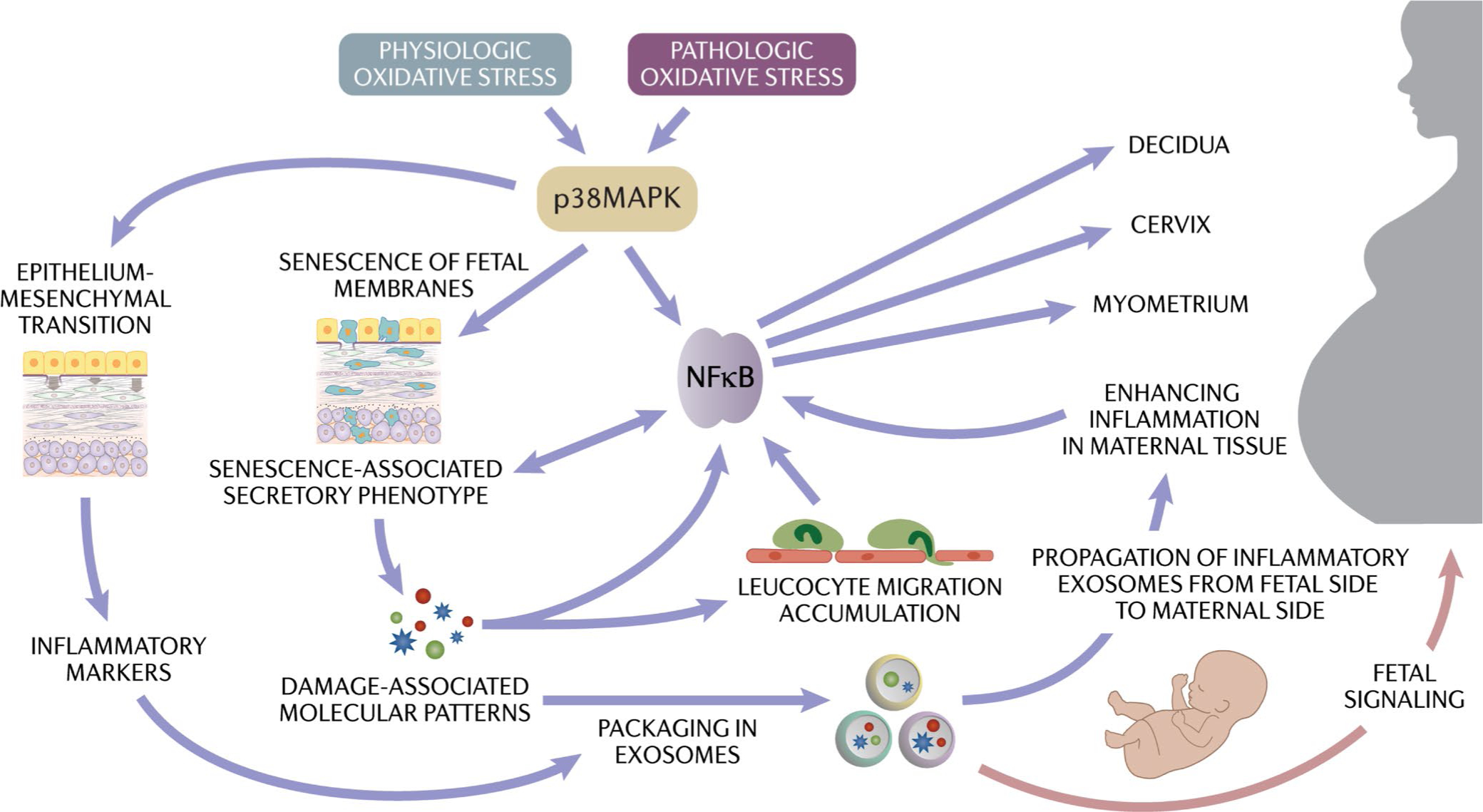
Human parturition at term and preterm is an inflammatory process synchronously executed by both fetomaternal tissues to transition them from a quiescent state t an active state of labor to ensure delivery. The initiators of the inflammatory signaling mechanism can be both maternal and fetal. The placental (fetal)-maternal immune and endocrine mediated homeostatic imbalances and inflammation are well reported. However, the fetal inflammatory response (FIR) theories initiated by the fetal membranes (amniochorion) at the choriodecidual interface are not well established. Although immune cell migration, activation, and production of proparturition cytokines to the fetal membranes are reported, cellular level events that can generate a unique set of inflammation are not well discussed. This review discusses derangements to fetal membrane cells (physiologically and pathologically at term and preterm, respectively) in response to both endogenous and exogenous factors to generate inflammatory signals. In addition, the mechanisms of inflammatory signal propagation (fetal signaling of parturition) and how these signals cause immune imbalances at the choriodecidual interface are discussed. In addition to maternal inflammation, this review projects FIR as an additional mediator of inflammatory overload required to promote parturition.

Keywords: EMT; aging; amniochorion; choriodecidua; exosomes; inflammation; premature rupture of the membranes; preterm birth; senescence; signaling.


Conflict of interest statement

CONFLICT OF INTEREST

Dr. Menon's research work was partly supported by ILIAS Biologics, Daejeon, S Korea between 2019–2021.

Figures

FIGURE 1
FIGURE 1
Pathways of preterm birth and current intervention strategies. During pregnancy, oxidative stress and inflammation generated in response to various risk factors lead to preterm labor-inducing pathways. Current interventions are designed primarily to reduce uterine contractions and cervical changes predisposing to preterm labor
FIGURE 2
FIGURE 2
Schematic representation of maintenance of the fetal membrane integrity and its disruption due to cellular transitions: The Amnion membrane is comprised of amnion epithelial cells (AEC) and amnion mesenchymal cells (AMC) separated by a Type 4 basement membrane collagen (BM) and extracellular matrix (ECM). During gestation, progesterone (P4) helps to maintain AEC epithelial state. AMCs, located in the extracellular matrix, express a fibroblastoid morphology. The ratio between AEC and AMC is 10:1 in normal gestation. EMT is mediated by TGF-β/TGF-β receptor-mediated mechanisms regulated by p38MAPK (middle panel; left). P4 and progesterone membrane receptor component (PGRMC2) mediated MET recycles AMC back to AEC to maintain tissue homeostasis and prevent accumulation of AMCs in the membrane extracellular matrix p38MAPK (middle panel; right). Oxidative stress causes increased activation of p38MAPK at term or preterm leading to increased EMT and a reduction in the function of progesterone as oxidative stress down-regulates PGRMC2 in AMCs. As the recycling of cells is stalled, AMC accumulates in the ECM and causes inflammatory changes (bottom panel). This figure was originally publsihed in Sci Signaling Richardson et al. reproduced here with permission
FIGURE 3
FIGURE 3
Senescence and paracrine signaling: A. Amnion epithelial (AEC), amnion mesenchymal (AMC), and chorion cells (CTC) (normal morphology under light microscopy), immunofluorescent staining of cytokeratin (CK)-18 (red), vimentin (green) staining show a meta-state of AEC, vimentin-positive AMC, and predominantly CK-18 positive (red) CTCs. Sensecence in AEC, AMC and CTC in response to oxidative stress is shown (blue staining cells) B. Infection, inflammation and other mechanisms of reactive oxygen species (ROS) increase can cause senescence of membrane cells. C. Senescence cause the release of senescence associated secretory phenotype (SASPs) and damage associated molecular pattern (DAMPs). SASPs and DAMPs are packaged inside the exosomes, which can cause paracrine signaling within the membrane cells. D. Senescence and inflammation increase within the fetal membrane cells and environment, leading to the propagation of exosomes to maternal decidua, myometrium, and cervix
FIGURE 4
FIGURE 4
Tracking fetal-derived exosomes and cells during pregnancy. A. Generation of transgenic mouse model containing TDTomato construct. A cre-reporter transgenic mouse model with membrane-targeted tdTomato (mT) A: A 2-color fluorescent Cre-reporter allele (mT/ mG construct) which has a membrane-targeted tandem dimer Tomato (mT) red fluorescent protein expressed in all cells and tissues, while enhanced green fluorescent protein (mG) is not expressed. Mating strategy to producing mT+expressing progeny. B. Immunohistology shows fetal exosomes (mT+, red staining), and flow cytometry data show 20% of maternal blood during gestation contains fetal (mT+) exosomes. C. Left panel: mT+cells are localized in various maternal organs. The placenta (fetal in origin) is expected to contain mT+in all the cells, as shown in the figure. mT+cells are seen in the uterus and even in maternal lungs. Right panel: Fetal Macrophage (φ) can be detected in maternal tissues. Confocal microscopy was used to colocalize mT expressing cells (red membrane fluorescence) with Mφ marker F4/80 (green membrane fluorescence) in the maternal cervix and uterus. 3D models were generated to confirm the expression of mT signal throughout the membrane of the cell. Scale bars represent 10 μm. See white arrows indicating mT signals surrounding cell membranes (n = 4) (these figures are reproduced with permission from Science family of journals/AAAS)
FIGURE 5
FIGURE 5
Determination of fetal neutrophils in the fetal membrane (FM) and the decidua: mT+neutrophils population was quantitated in tissue homogenates from the fetal membranes and decidua after an LPS challenge of animals (red bars). A. In the fetal membranes, the total neutrophis (maternal+fetal) was increased significantly after an LPS challenge, whereas mT-(maternal) was not changed. Fetal (mT+) was significantly higher in the membranes after the LPS challenge compared to PBS (blue bars). B. In the decidua, LPS challenge decreased neutrophils slightly compared to total (regardless of fetal vs. maternal)
FIGURE 6
FIGURE 6
A. Representation of fetal membranes. The chorio-decidual region is shown in the box with broken lines. B. Chorion trophoblast cells (CTC) in culture for 48 h (left), treated with exosomes from amnion epithelial cells (AEC) grown under normal cell culture conditions (middle), or treated with cigarette smoke extract (CSE) treated oxidatively stressed (OS) AEC-derived exosomes. Exosomes from OS stressed AECs transitioned CTCs to CMCs. C. NETosis induction by exosomes: Representative images from a study using neutrophils from healthy pregnant subjects. Control cells do not show NETosis. Phorbol 12-myristate 13-acetate (PMA) and CSE-treated AEC-derived exosomes induced a higher number of NETosis compared to control AEC-derived exosomes. D. Flow cytometry. Decidual CD45+ cells were examined after treatment with senescent AEC exosomes. CD14, CD3, and CD66b antibodies were tested for NK cells, neutrophils, and monocytes. Compared to normal AEC-derived exosomes, CSE-treated AEC exosomes increased CD66b+ cells, whereas no change in CD14+ and CD3+ cells were seen. Figure 6A is a drawing by Dr. Lauren Richardson (Assistant Professor, The University of Texas Medical Branch at Galveston)
FIGURE 7
FIGURE 7
Propagation of exosomes and expected changes at the choriodecidual interface. Chorion trophoblast cells are rich sources of progesterone and IL-10 with anti-inflammatory properties and help to maintain choriodecidual homeostasis. Fetal cell exosomes generated during pregnancy can carry fetal cellular signals (shown in green circles) (top panel); however, they are natural and physiologic responses and insufficient to change the choriodecidual interface's homeostasis. In response to intrauterine infection or inflammation, oxidative stress generated can accelerate membrane cell EMT and senescence. These pathologic events can generate inflammatory cargo enriched exosomes (shown in red circles) (bottom panel) that can increase TNF-α production decrease in P4, and IL-10 release. Immune cell infiltration in response to this event can bring neutrophils to the fetal membranes that can cause NETosis. These inflammatory changes can compromise the integrity of the choriodecidual interface
FIGURE 8
FIGURE 8
Physiologic (term) and pathologic (preterm) activation of oxidative stress and inflammation and the cascade of events resulting in parturition. Generation of the fetal inflammatory response (FIR) due to the cellular level changes can generate exosomes packaged in signalers that can transition quiescent maternal uterine tissues to an active state of labor (fetal signaling generated by changes to the fetal membrane cells)

References

    1. Romero R, Dey SK, Fisher SJ. Preterm labor: one syndrome, many causes. Science. 2014;345(6198):760–765. - PMC - PubMed
    1. Tal R, Taylor HS. Endocrinology of pregnancy. In: Feingold KR, Anawalt B, Boyce A, et al., Endotext. MDText.com, Inc.; 2000.
    1. Rokas A, Mesiano S, Tamam O, LaBella A, Zhang G, Muglia L. Developing a theoretical evolutionary framework to solve the mystery of parturition initiation. Elife. 2020;9. - PMC - PubMed
    1. Smith R Alterations in the hypothalamic pituitary adrenal axis during pregnancy and the placental clock that determines the length of parturition. J Reprod Immunol. 1998;39(1–2):215–220. - PubMed
    1. Menon R, Bonney EA, Condon J, Mesiano S, Taylor RN. Novel concepts on pregnancy clocks and alarms: redundancy and synergy in human parturition. Hum Reprod Update. 2016;22(5):535–560. - PMC - PubMed

Publication types