. 2022 Dec;60(1):609-620.
doi: 10.1080/13880209.2022.2043392.

Banxia-Houpu decoction diminishes iron toxicity damage in heart induced by chronic intermittent hypoxia

Affiliations

Banxia-Houpu decoction diminishes iron toxicity damage in heart induced by chronic intermittent hypoxia

Ji-Xian Song et al. Pharm Biol. 2022 Dec.

Abstract

Context: Obstructive sleep apnoea (OSA) causes chronic intermittent hypoxia (CIH), which results in mitochondrial dysfunction and generates reactive oxygen species (ROS) in the heart. Excessive free iron could accelerate oxidative damage, which may be involved in this process. Banxia-Houpu decoction (BHD) was reported to improve the apnoea hypopnoea index in OSA patients, but the specific mechanism was still unclear.

Objective: To investigate whether BHD could reduce CIH-induced heart damage by regulating iron metabolism and mitochondrial function.

Materials and methods: C57BL/6N mice were randomly divided into control, CIH and BHD groups. Mice were exposed to CIH (21 - 5% O2, 20 times/h, 8 h/d) and administered BHD (3.51, 7.01 and 14.02 g/kg, intragastrically) for 21 d. Cardiac and mitochondrial function, iron levels, apoptosis and mitophagy were determined.

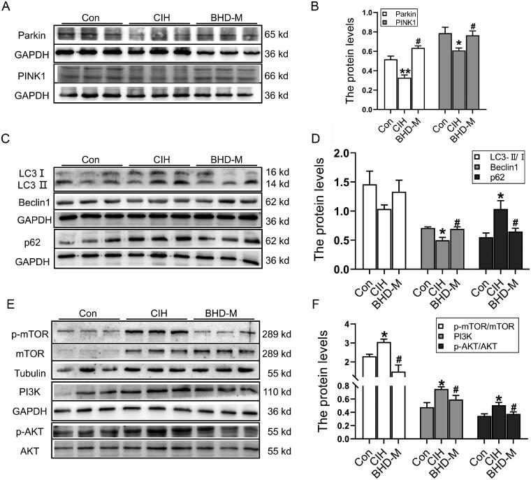
Results: BHD (7.01 g/kg) significantly improved cardiac dysfunction, pathological change and mitochondrial structure induced by CIH. BHD increased the Bcl-2/Bax ratio (1.4-fold) and inhibited caspase 3 cleavage in CIH mice (0.45-fold). BHD activated mitophagy by upregulating Parkin (1.94-fold) and PINK1 (1.26-fold), inhibiting the PI3K-AKT-mTOR pathway. BHD suppressed ROS generation by decreasing NOX2 (0.59-fold) and 4-HNE (0.83-fold). BHD reduced the total iron in myocardial cells (0.72-fold) and mitochondrial iron by downregulating Mfrn2 (0.81-fold) and MtFt (0.78-fold) proteins, and upregulating ABCB8 protein (1.33-fold). Rosmarinic acid, the main component of Perilla Leaf in BHD, was able to react with Fe2+ and Fe3+ in vitro.

Discussion and conclusions: These findings encourage the use of BHD to resist cardiovascular injury and provide the theoretical basis for clinical treatment in OSA patients.

Keywords: Obstructive sleep apnoea; cardiac damage; mitochondrial disfunction; rosmarinic acid.


Conflict of interest statement

No potential conflict of interest was reported by the author(s).

Figures

Figure 1.
Figure 1.
Quantitative analysis of components in the aqueous of Banxia Houpu decoction by LC-MS/MS. (A,B) The magnolol of standards (St) and BHD. (C,D) The honokiol of standards (St) and BHD. (E,F) The succinic acid of standards (St) and BHD. (G,H) The rosmarinic acid of standards (St) and BHD. (I,J) The 6-gingerol of standards (St) and BHD.
Figure 2.
Figure 2.
Cardiac dysfunction induced by CIH exposure in mice. (A) M-model echocardiography in mice (n = 6). (B) The ejection fraction (EF) of the left ventricle (n = 6). (C) The fractional shortening (FS) (n = 6). (D) The left ventricular end-systolic volume (LVESV, n = 6). (E) The left ventricular end-diastolic volume (LVEDV, n = 6). (F) The velocity ratio of the E peak to the A peak in the cardiac mitral valve (MV E/A) (n = 6). (G,H) The H&E staining and Masson’s trichrome staining of the Con, CIH, BHD-L, BHD-M and BHD-H groups (scale bar = 20 μm, n = 3). The data are presented as the mean ± SEM. **p < 0.01 vs. Con group. #p < 0.05, ##p < 0.01 vs. CIH group.
Figure 3.
Figure 3.
Mitochondrial damage and mitochondrial pathway-dependent apoptosis in the hearts of CIH mice. (A) The TEM images of mitochondria in the heart. (B,C) The expression of Opa1 and Drp1 proteins in heart tissue by western blot. (D) The TUNEL staining of heart tissue (scale bar = 12.5 μm, n = 3). (E) The total number of apoptotic cells as shown in panel D. (F,G) The expression of Bcl-2/Bax and cleaved-caspase 3/pro-caspase 3 proteins in heart tissue by western blot. The data are presented as the mean ± SEM, n = 3. *p < 0.05, **p < 0.01 vs. Con group. #p < 0.05, ##p < 0.01 vs. BHD-M group.
Figure 4.
Figure 4.
Cardiac autophagy and the PI3K/AKT/mTOR signalling pathway after exposure to CIH. (A,B) The expression of Parkin and PINK1 proteins in heart tissue by western blot. (C,D) The expression of LC3B, Beclin-1, p62 proteins in heart tissue by western blot. (E,F) The expression of PI3K/AKT/mTOR signalling pathway in heart tissue by western blot. The data are presented as the mean ± SEM, n = 3. *p < 0.05, **p < 0.01 vs. Con group. #p < 0.05 vs. BHD-M group.
Figure 5.
Figure 5.
Oxidative stress levels in cardiac tissue subjected to CIH. (A) DHE staining in heart tissue (scale bar = 25 μm, n = 3). (B) The mean fluorescence intensity of DHE as shown in panel A. (C,D) The expression and statistics of NOX2 and 4-HNE protein levels. The results are presented as the mean ± SEM, n = 3. **p < 0.01 vs. Con group. #p < 0.05, ##p < 0.01 vs. BHD-M group.
Figure 6.
Figure 6.
Iron content and iron-related transport proteins in cardiac tissue and mitochondria during CIH. (A) Perls’ staining of heart tissue (scale bar = 25 μm). (B) The mean density of Fe content as shown in panel A. (C,D) The expression and statistics of the iron-related transport proteins, TfR1, DMT1(−ire), DMT1(+ire) and FPN1. (E,F) The expression and statistics of iron transporter-associated proteins in mitochondria, Mfrn2, MtFt and ABCB8. The results are presented as the mean ± SEM, n = 3. ∗p < 0.05, **p < 0.01 vs. Con group. #p < 0.05 vs. BHD-M group.
Figure 7.
Figure 7.
The main component of BHD could specifically chelate iron. The λ absorption of (A) Magnolol, (B) Honokiol, (C) Succinic acid, (D) 6-Gingerol, (E) Rosmarinic acid with Fe2+ and Fe3+, respectively. (F) The enlarged images as shown in panel E.
Figure 8.
Figure 8.
A schematic graph of the proposed cardioprotective mechanism of BHD when exposed to CIH. BHD reduced iron deposition, particularly in mitochondria, by downregulating TfR1, DMT1, Mfrn2 and MtFt, and upregulating FPN1 and ABCB8 expression, and then inhibiting the high level of ROS. BHD enhanced mitochondrial autophagy via the PI3K/AKT/mTOR signalling pathway to relieve mitochondrial dysfunction and mitochondrial-dependent apoptosis to exert cardioprotective effects.

Similar articles

Cited by

References

    1. An JR, Zhao YS, Luo LF, Guan P, Tan M, Ji ES.. 2020. Huperzine A, reduces brain iron overload and alleviates cognitive deficit in mice exposed to chronic intermittent hypoxia. Life Sci. 250:117573–10. - PubMed
    1. Anderson ER, Shah YM.. 2013. Iron homeostasis in the liver. Compr Physiol. 3(1):315–330. - PMC - PubMed
    1. Ban T, Ishihara T, Kohno H, Saita S, Ichimura A, Maenaka K, Oka T, Mihara K, Ishihara N.. 2017. Molecular basis of selective mitochondrial fusion by heterotypic action between OPA1 and cardiolipin. Nat Cell Biol. 19(7):856–863. - PubMed
    1. Benjafield AV, Ayas NT, Eastwood PR, Heinzer R, Ip MSM, Morrell MJ, Nunez CM, Patel SR, Penzel T, Pepin JL, et al. . 2019. Estimation of the global prevalence and burden of obstructive sleep apnoea: a literature-based analysis. Lancet Respir Med. 7(8):687–698. - PMC - PubMed
    1. Brentnall M, Rodriguez-Menocal L, De Guevara RL, Cepero E, Boise LH.. 2013. Caspase-9, caspase-3 and caspase-7 have distinct roles during intrinsic apoptosis. BMC Cell Biol. 14:32–39. - PMC - PubMed

MeSH terms