. 2022 Apr:93:106932.
doi: 10.1016/j.ijscr.2022.106932. Epub 2022 Mar 9.

Surgical management of large, connected perineal and pelvic epidermal inclusion cysts mimicking a dumbbell-shaped lesion in an adult male

Affiliations

Surgical management of large, connected perineal and pelvic epidermal inclusion cysts mimicking a dumbbell-shaped lesion in an adult male

Dakota T Thompson et al. Int J Surg Case Rep. 2022 Apr.

Abstract

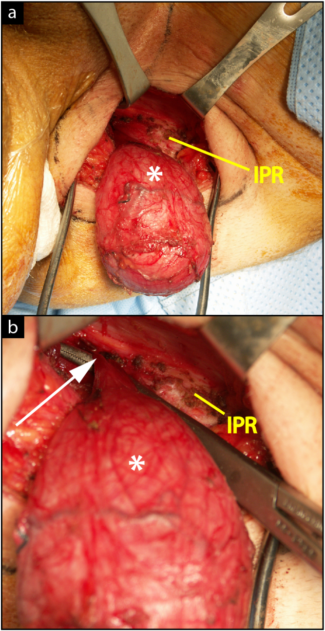
Introduction and importance: Epidermal inclusion cysts are a common benign finding, and they are predominantly asymptomatic. They can rarely form in the pelvis or abdomen, however, and may cause symptoms secondary to mass effect. This case highlights management of an anterectal epidermal inclusion cyst connected to the perineal cyst, mimicking a dumbbell-shaped lesion, found in a male.

Case presentation: This is a unique case of a 21-year-old Caucasian male with a palpable perineal mass, lower extremity hypoesthesia, and constipation who was found to have a complex-shaped cyst on computed tomography and magnetic resonance imaging. This was ultimately managed with a two-stage perineal and transabdominal resection.

Clinical discussion: This case highlights that perineal epidermal inclusion cysts may have pelvic extension, especially in patients with additional new-onset neurologic, gastrointestinal, or urologic symptoms. These symptoms should completely resolve after resection. Additionally, resection is recommended to prevent complications including malignant degeneration and fistulization.

Conclusion: This is the first reported case of an anterectal, epidermal inclusion cyst connected to a perineal cyst found in a male. Perineal and pelvic cysts may be synchronous and may be connected through the pudendal canal. These masses can be safely removed via a combined perineal and transabdominal resection. The connecting portion of lesions that have both pelvic and perineal components should be meticulously identified and dissected because even a thin, patent segment - if left unresected - may result in lesion recurrence.

Keywords: Case report; Dumbbell-shaped; Epidermal inclusion cyst; Perineal cyst; Rectovesical pouch.


Conflict of interest statement

The authors have no conflicts of interest to declare.

Figures

Fig. 1
Fig. 1
Preoperative contrast-enhanced CT showing well-defined unilocular hypodense mass with a thin wall anterior to the rectum. a) Axial CT scan of pelvis showing pelvic mass (yellow outline) next to pudendal canal (PC, red outline), bladder (BL), iliacus muscle (IL), acetabulum (A), gluteus maximus muscle (GM), obturator internus muscle (OI), external iliac artery (EIA), piriformis muscle (PI), sacrum (S), and rectum (*). b) Axial CT scan of more caudal section highlighting perineal portion of mass (blue outline), semimembranosus muscle (SM), tensor fasciae latae muscle (TFL), vastus lateralis muscle (VL), adductor longus muscle (AL), rectus femoris muscle (RF), and gluteus maximus muscle (GM). c) Coronal CT scan showing both pelvic (yellow outline) and perineal (blue outline) portions of mass, iliac crest (IL), sacrum (S), ischium (IS), gluteus maximus muscle (GM), and rectum (*). d) Sagittal CT scan showing pelvic (yellow outline) and perineal (blue outline) portions of mass, bladder (BL), and sacrum (S).
Fig. 2
Fig. 2
Preoperative contrast-enhanced T1- and T2-weighted MRI showing pelvic mass. a) Axial T1-weighted MRI showing hypointense homogeneous cystic lesion (yellow outline) without enhancement displacing the rectum (*) and next to the pudendal canal (PC, red outline), rectus abdominis (R), iliacus muscle (IL), gluteus maximus muscle (GM), obturator internus muscle (OI), and acetabulum (A). b) Axial T2-weighted MRI showing homogeneous hyperintense lesion (yellow outline).
Fig. 3
Fig. 3
Intraoperative findings from resection of the perineal portion of the dumbbell-shaped epidermal inclusion cyst. a) The cyst (*) was able to be completely mobilized from the subcutaneous tissue of the left buttock adjacent to the inferior pubic rami (IPR). b) Finding of fibrous band (white arrow) connected to cyst (*) near the inferior pubic rami (IPR).
Fig. 4
Fig. 4
Additional intraoperative findings and histologic findings from resection of the perineal portion of the dumbbell-shaped epidermal inclusion cyst. a) Perineal cyst containing desquamated material. The obliterated band extending through the pudendal canal is marked with an arrow. b) Microscopy of the cyst wall (200× magnification) with Hematoxylin and Eosin Staining demonstrating keratinizing-stratified squamous epithelial lining (arrow) and a prominent granular layer (image courtesy of Robert A. Robinson, MD).

References

    1. Fakhir B., Mamouni N., Bouramdane N., et al. A rare case of a giant pelvic retroperitoneal epidermoid cyst. Libyan J. Med. Jun 2009;4(2):61. doi: 10.4176/090210. - DOI - PMC - PubMed
    1. Hobson K.G., Ghaemmaghami V., Roe J.P., Goodnight J.E., Khatri V.P. Tumors of the retrorectal space. Dis. Colon Rectum. Oct 2005;48(10):1964–1974. doi: 10.1007/s10350-005-0122-9. - DOI - PubMed
    1. Jain P., Pal D.K. Pelvic epidermoid cyst: a rare cause of lower urinary tract symptoms. BMJ Case Rep. May 2018;2018 doi: 10.1136/bcr-2017-223258. - DOI - PMC - PubMed
    1. Palanivelu C., Rangarajan M., Senthilkumar R., Madankumar M.V., Annapoorni S. Laparoscopic and perineal excision of an infected "dumb-bell" shaped retrorectal epidermoid cyst. J. Laparoendosc. Adv. Surg. Tech. A. Feb 2008;18(1):88–92. doi: 10.1089/lap.2007.0010. - DOI - PubMed
    1. Agha R.A., Franchi T., Sohrabi C., Mathew G., Kerwan A., Group S The SCARE 2020 guideline: updating consensus Surgical CAse REport (SCARE) guidelines. Int. J. Surg. Dec 2020;84:226–230. doi: 10.1016/j.ijsu.2020.10.034. - DOI - PubMed