

Bowen's Disease
- PMID: 35287414
- PMCID: PMC8917478
- DOI: 10.4103/idoj.idoj_257_21
Bowen's Disease
Abstract
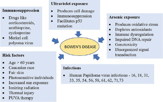
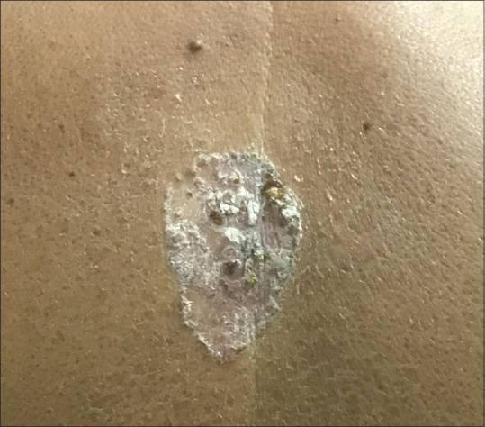
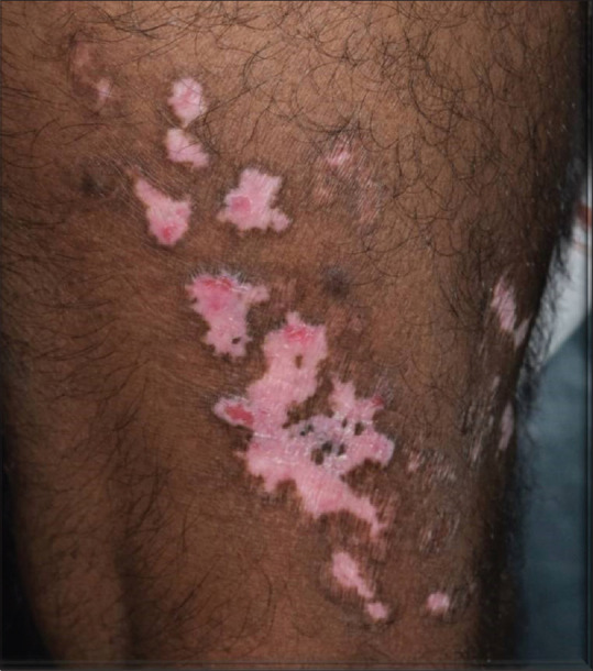
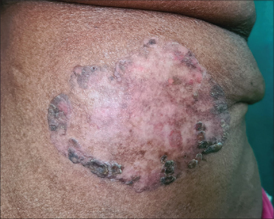
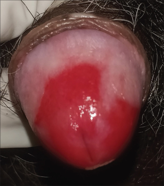
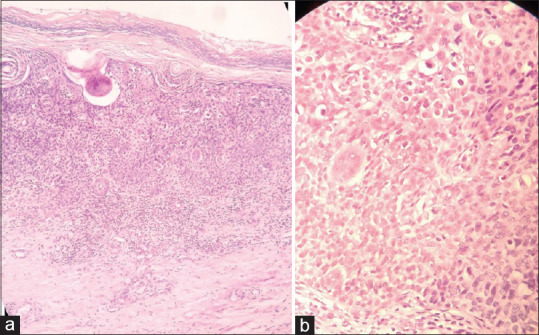
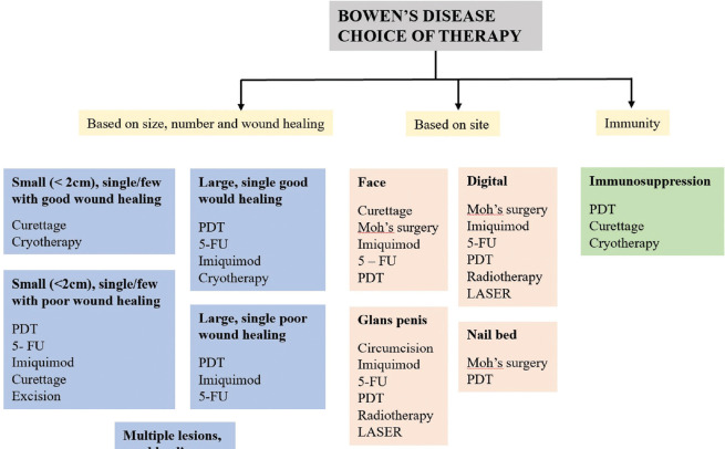
Bowen's disease (BD) is an in-situ squamous cell carcinoma of epidermis. The etiology of BD is multifactorial with high incidence among Caucasians. BD is common in photo-exposed areas of skin, but other sites can also be involved. Lesions are usually solitary. The morphology of BD differs based on age of the lesion, site of origin, and the degree of keratinization. BD is considered as the "lull before the storm," which precedes an overt squamous cell carcinoma. Histopathology is the gold standard diagnostic modality to confirm the diagnosis. Immunohistochemistry, dermoscopy, and reflectance confocal microscopy are the adjuvant modalities used in the diagnosis of BD. The treatment depends on various factors like site, size, immune status, patient's age, esthetic outcome, etc. The available therapeutic modalities include topical chemotherapy, surgical modalities, light-based modalities, and destructive therapies. It requires a combined effort of dermatologist, oncosurgeon, and plastic surgeon to plan and execute the management in various presentations of BD.
Keywords: Bowen's disease; erythroplasia of Queyrat; squamous cell carcinoma.
Copyright: © 2022 Indian Dermatology Online Journal.
Conflict of interest statement
There are no conflicts of interest.
Figures
References
-
- Heppt MV, Schlager G, Berking C. Epithelial precancerous lesions. In: Kang S, Amagai M, Bruckner AL, Enk AH, Margolis DJ, McMichael AJ, et al., editors. Fitzpatrick's Dermatology in General Medicine. 9th ed. New York: Mcgraw-Hill; 2019. pp. 1857–83.
-
- Arlette JP, Trotter MJ. Squamous cell carcinoma in situ of the skin: History, presentation, biology and treatment. Australas J Dermatol. 2004;45:1–9. - PubMed
-
- Bowen JT. Precancerous dermatosis. J Cutan Dis. 1912;30:241. - PubMed
-
- Bernhard JD, Elliot AD. A letter from darier to bowen on the naming of Bowen's disease. Arch Dermatol. 1983;119:261–2. - PubMed
-
- Darier J. La dermatose precancereuse de Bowen-dyskeratose lentiularie et en disques. Ann Dermatologie. 1914;5:449–71.
Publication types
LinkOut - more resources
Full Text Sources
