. 2022 Mar 14;10(1):33.
doi: 10.1186/s40478-022-01334-7.

TMEM106B deficiency impairs cerebellar myelination and synaptic integrity with Purkinje cell loss

Affiliations

TMEM106B deficiency impairs cerebellar myelination and synaptic integrity with Purkinje cell loss

Tuancheng Feng et al. Acta Neuropathol Commun. .

Abstract

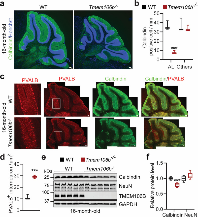
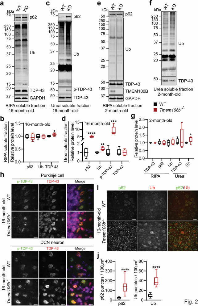
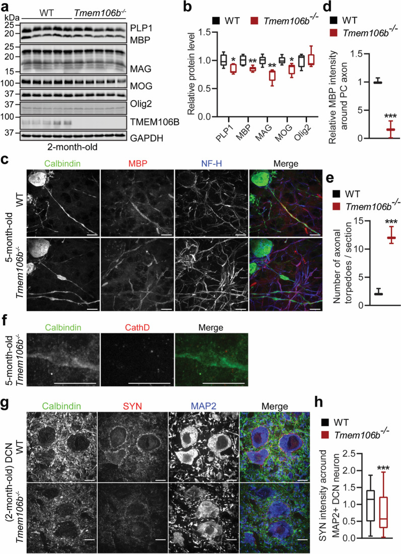
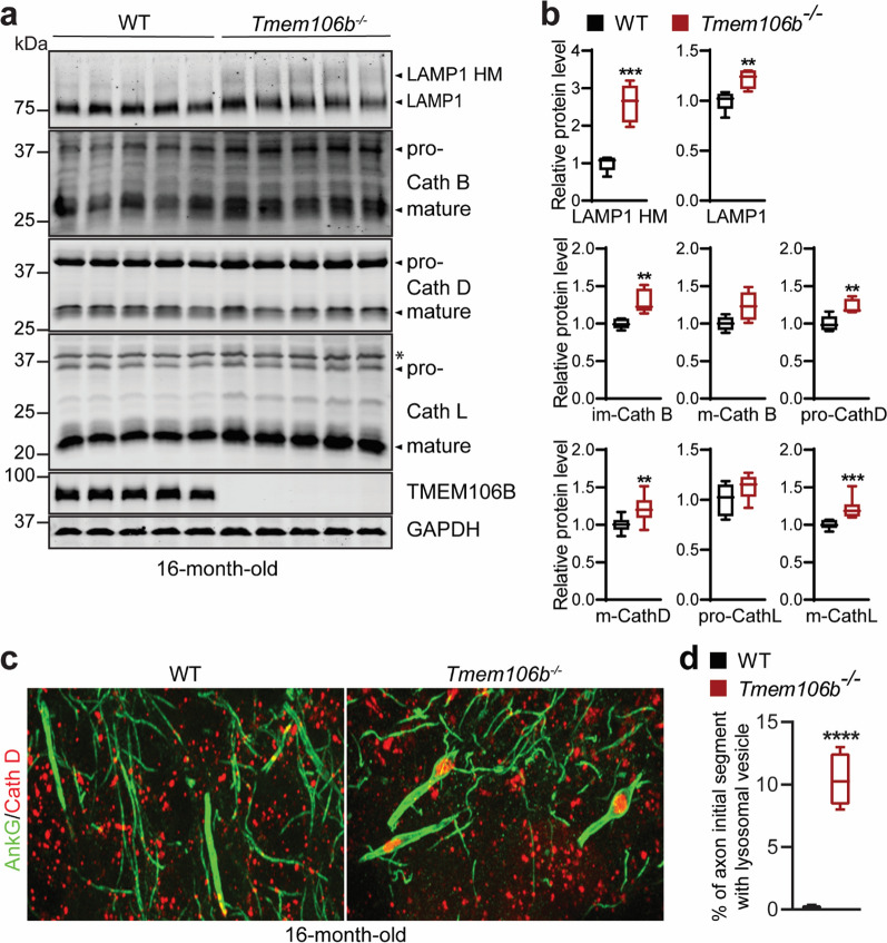
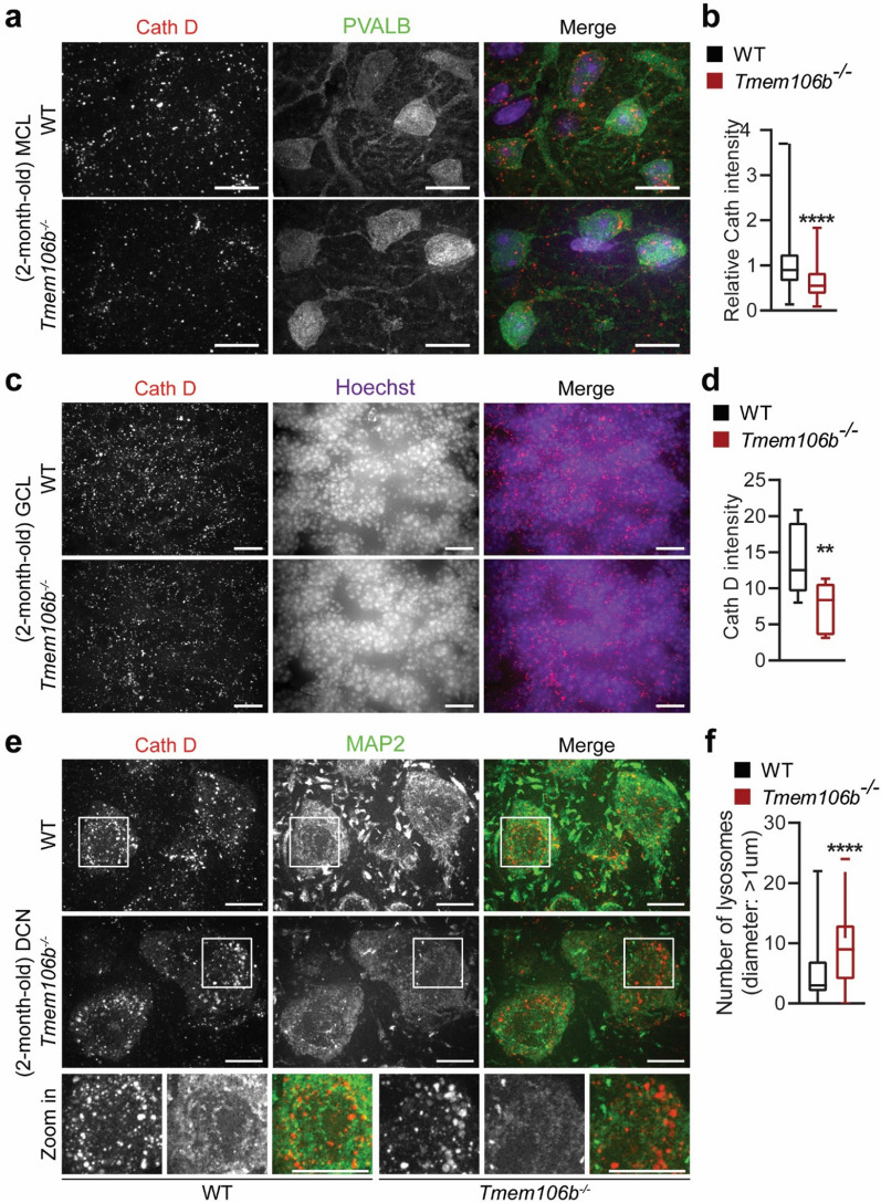
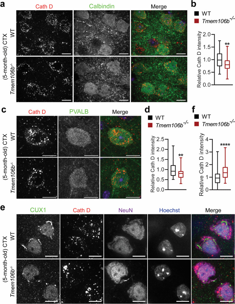
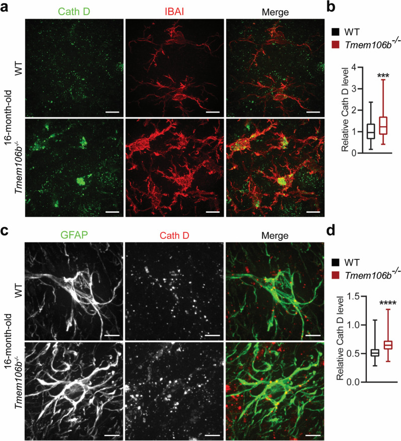
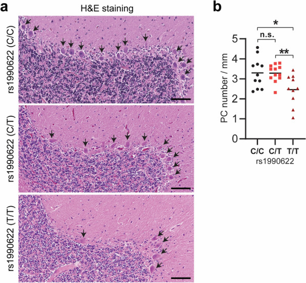
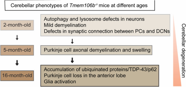
TMEM106B, a type II lysosomal transmembrane protein, has recently been associated with brain aging, hypomyelinating leukodystrophy, frontotemporal lobar degeneration (FTLD) and several other brain disorders. TMEM106B is critical for proper lysosomal function and TMEM106B deficiency leads to myelination defects, FTLD related pathology, and motor coordination deficits in mice. However, the physiological and pathological functions of TMEM106B in the brain are still not well understood. In this study, we investigate the role of TMEM106B in the cerebellum, dysfunction of which has been associated with FTLD and other brain disorders. We found that TMEM106B is ubiquitously expressed in neurons in the cerebellum, with the highest levels in the Purkinje neurons. Aged TMEM106B-deficient mice show significant loss of Purkinje neurons specifically in the anterior lobe of the cerebellum. Increased microglia and astrocyte activation, as well as an accumulation of ubiquitinated proteins, p62 and TDP-43 were also detected in the cerebellum of aged TMEM106B deficient mice. In the young mice, myelination defects and a significant loss of synapses between Purkinje and deep cerebellar nuclei neurons were observed. Interestingly, TMEM106B deficiency causes distinct lysosomal phenotypes in different types of neurons and glia in the cerebellum and frontal cortex. In humans, TMEM106B rs1990622 risk allele (T/T) is associated with increased Purkinje neuron loss. Taken together, our studies support that TMEM106B regulates lysosomal function in a cell-type-specific manner and TMEM106B is critical for maintaining synaptic integrity and neural functions in the cerebellum.

Keywords: Cerebellum; FTLD; Lysosome; Myelination; TMEM106B.


Conflict of interest statement

The authors declare that they have no competing interests.

Figures

Fig. 1
Fig. 1
Degeneration of Purkinje cells in aged Tmem106b−/− mice. a, b Cerebellar sections from 16‐month‐old WT and Tmem106b−/− mice were stained with anti-calbindin antibody and Hoechst. Purkinje cell numbers in the anterior lobe (AL) and other regions (Others) were quantified in b. Scale bar = 100 µm. n = 3–4, ***, p < 0.001, unpaired t-test. c, d Cerebellar sections from 16‐month‐old WT and Tmem106b−/− mice were immuno-stained with antibodies of calbindin and parvalbumin (PVALB). Zoom-in images show PVALB-positive interneurons in the MCL adjacent to the region with Purkinje cell loss. The number of PVALB-positive interneurons in the molecular layer of the anterior lobe (AL) was quantified in d. Scale bar = 100 µm. n = 3, ***, p < 0.001, unpaired t-test. e, f Western blot analysis of the protein level of Calbindin, NeuN, and TMEM106B in 16-month-old WT and Tmem106b−/− cerebellar lysates. GAPDH was used as an internal control. Relative protein levels were quantified in f. n = 5, ***, p < 0.001, unpaired t-test
Fig. 2
Fig. 2
ALS/FTLD related pathological changes in the cerebellum of aged Tmem106b−/− mice. ad Western blot analysis of p62, ubiquitin (Ub), TDP‐43, and p‐TDP-43 in RIPA—(a, b) and urea—(c, d) soluble fractions from the cerebellum of 16‐month‐old WT and Tmem106b−/− mice. Protein levels were quantified and normalized to GAPDH. n = 5, *, p < 0.05; **, p < 0.01, ***, p < 0.001, ****, p < 0.0001, unpaired t-test. eg Western blot analysis of p62, ubiquitin (Ub), and TDP‐43 in RIPA—(e, g) and urea—(f, g) soluble fractions from the cerebellum of 2-month‐old WT and Tmem106b−/− mice. Protein levels were quantified and normalized to GAPDH. n = 5, *, p < 0.05, unpaired t-test. h Immunostaining of TDP‐43 and p‐TDP-43 in the cerebellar sections from 16‐month‐old WT and Tmem106b−/− mice. Representative images from the Purkinje cell layer and deep cerebellar nuclei (DCN) are shown. i, j Immunostaining of p62 and ubiquitin (Ub) in the cerebellar sections from 16‐month‐old WT and Tmem106b−/− mice. The number of p62 or Ub positive puncta was quantified in (j). n = 4. ****, p < 0.0001, unpaired t-test
Fig. 3
Fig. 3
Myelination defects, axonal degeneration of Purkinje cells and disruption of cerebellar cortico-nuclear connection in young Tmem106b−/− mice. a, b Western blot analysis of myelin proteins and GAPDH in 2-month-old WT and Tmem106b−/− cerebellar lysates. Protein levels were quantified and normalized to GAPDH in b. n = 5, *, p < 0.05, **, p < 0.01, unpaired t-test. ce Cerebellar sections from 5‐month‐old WT and Tmem106b−/− mice were co-stained with anti-calbindin, myelin basic protein (MBP), and NF-H antibodies. MBP intensity around Purkinje cell axon and the number of giant torpedos in the axon of Purkinje cells were quantified in d and e, respectively. Scale bar = 10 µm. n = 3–4, ***, p < 0.001, unpaired t-test. f Cerebellar sections from 5‐month‐old Tmem106b−/− mice were co-stained with anti-calbindin and Cath D antibodies. Scale bar = 10 µm. g, h Cerebellar sections from 2‐month‐old WT and Tmem106b−/− mice were immunostained with antibodies of calbindin, synaptophysin (SYN, presynaptic marker), and MAP2. The intensity of SYN around MAP2-positive soma in the DCN region was quantified in h. Scale bar = 10 µm. n = 3, ***, p < 0.001, non-parametric test (Mann Whitney test)
Fig. 4
Fig. 4
Lysosome and lysosomal trafficking defects in the cerebellum of aged Tmem106b−/− mice. a, b Western blot analysis of lysosomal proteins and GAPDH in 16-month-old WT and Tmem106b−/− cerebellar lysates. Protein levels were quantified and normalized to GAPDH in b. HM: high molecular weight. n = 5, **, p < 0.01, ***, p < 0.001, unpaired t-test. Asterisk indicates non‐specific bands. c, d Immunostaining of AnkG and Cath D in the cerebellar sections from 16‐month‐old WT and Tmem106b−/− mice. Representative images from the Purkinje cell layer are shown. The percentage of axon initial segments (AISs) with Cath D-positive lysosomal vesicles were quantified in d. n = 3. ****, p < 0.0001, unpaired t-test
Fig. 5
Fig. 5
Distinct lysosomal phenotypes among different types of neurons in the cerebellum of young Tmem106b−/− mice. a, b Immunostaining of PVALB and Cathepsin D (Cath D) in cerebellar sections from 2-month‐old WT and Tmem106b−/− mice. Representative images from the molecular cell layer are shown. The intensity of Cath D in PVALB-positive interneurons was quantified in b. n = 3. ****, p < 0.0001, unpaired t-test. Scale bar = 10 μm. c, d Immunostaining of Cathepsin D (Cath D) and Hoechst in cerebellar sections from 2-month‐old WT and Tmem106b−/− mice, and images were captured from granule cell layer. The intensity of Cath D in the granule cell layer was quantified in d. n = 3. **, p < 0.001, unpaired t-test. Scale bar = 10 μm. e, f Immunostaining of Cathepsin D (Cath D) and MAP2 in cerebellar sections from 2-month‐old WT and Tmem106b−/− mice. Representative images from the DCN region are shown. The intensity of Cath D in MAP2-positive DCN neurons was quantified in f. Zoom-in image shows the enlarged lysosomes in 2-month‐old Tmem106b−/− mice compared with WT mice. n = 3. ****, p < 0.0001, unpaired t-test. Scale bar = 10 μm
Fig. 6
Fig. 6
Distinct lysosomal phenotypes among different types of neurons in the cortex (CTX) in young Tmem106b−/− mice. a, b Immunostaining of Calbindin and Cath D in brain sections from 5-month‐old WT and Tmem106b−/− mice, and images were captured from the frontal cortex. The intensity of Cath D in Calbindin-positive neurons was quantified in b. n = 3. **, p < 0.01, unpaired t-test. Scale bar = 10 μm. c, d Immunostaining of PVALB and Cathepsin D (Cath D) in brain sections from 5-month‐old WT and Tmem106b−/− mice. Representative images from the frontal cortex are shown. The intensity of Cath D in PVALB-positive interneurons was quantified in d. n = 3. **, p < 0.01, unpaired t-test. Scale bar = 10 μm. e, f Immunostaining of CUX1, Cath D, and NeuN in brain sections from 5-month‐old WT and Tmem106b−/− mice. Representative images from the frontal cortex are shown. The intensity of Cath D in CUX1-positive excitatory neuron was quantified in f. n = 3. ****, p < 0.0001, unpaired t-test. Scale bar = 10 μm
Fig. 7
Fig. 7
Lysosome defects in glial cells in the cerebellum of aged Tmem106b−/− mice. a, b Cerebellar sections from 16-month‐old WT and Tmem106b−/− mice were immunostained with IBA1 and Cath D antibodies. Representative images from the DCN region are shown. The intensity of Cath D in IBA1-positive microglia was quantified in b. n = 3. ***, p < 0.001, unpaired t-test. Scale bar = 10 μm. c, d Cerebellar sections from 16-month‐old WT and Tmem106b−/− mice were immunostained with GFAP and Cath D antibodies. Representative images from the DCN region are shown. The intensity of Cath D in GFAP-positive astrocytes was quantified in d. n = 3. ****, p < 0.0001, unpaired t-test. Scale bar = 10 μm
Fig. 8
Fig. 8
TMEM106B rs1990622 risk allele is associated with Purkinje cell loss in humans. a H&E staining was performed in human cerebellum sections from cohorts with different TMEM106B rs1990622 genotypes (C/C, C/T, and T/T). Representative images from each genotype are shown. b Number of Purkinje cells was counted in the cerebellar section, and the density of Purkinje cells along the Purkinje cell layer was calculated. n = 10–13 for each genotype, n.s., not significant, *, p < 0.05, **, p < 0.001, unpaired t-test. Scale bar = 100 µm
Fig. 9
Fig. 9
A diagram summarizing cerebellar pathologies in Tmem106b−/− mice with age. At 2 months of age, Tmem106b−/− young mice exhibit lysosome and myelination defects, and a significant loss of synapses between Purkinje and deep cerebellar nuclei neurons. Purkinje cell axonal demyelination and swelling are observed at 5 months of age. Significant loss of Purkinje neurons specifically in the anterior lobe of the cerebellum are observed in 16-month of age, accompanied by increased microglia and astrocyte activation, and the accumulation of ubiquitinated proteins, p62, and TDP-43

Similar articles

Cited by

References

    1. Adamaszek M, D'Agata F, Ferrucci R, Habas C, Keulen S, Kirkby KC, Leggio M, Marien P, Molinari M, Moulton E, Orsi L, Van Overwalle F, Papadelis C, Priori A, Sacchetti B, Schutter DJ, Styliadis C, Verhoeven J. Consensus paper: cerebellum and emotion. Cerebellum. 2017;16:552–576. doi: 10.1007/s12311-016-0815-8. - DOI - PubMed
    1. Bharti K, Khan M, Beaulieu C, Graham SJ, Briemberg H, Frayne R, Genge A, Korngut L, Zinman L, Kalra S, Canadian ALSNC. Involvement of the dentate nucleus in the pathophysiology of amyotrophic lateral sclerosis: a multi-center and multi-modal neuroimaging study. Neuroimage Clin. 2020;28:102385. doi: 10.1016/j.nicl.2020.102385. - DOI - PMC - PubMed
    1. Bocchetta M, Todd EG, Peakman G, Cash DM, Convery RS, Russell LL, Thomas DL, Eugenio Iglesias J, van Swieten JC, Jiskoot LC, Seelaar H, Borroni B, Galimberti D, Sanchez-Valle R, Laforce R, Moreno F, Synofzik M, Graff C, Masellis M, Carmela Tartaglia M, Rowe JB, Vandenberghe R, Finger E, Tagliavini F, de Mendonca A, Santana I, Butler CR, Ducharme S, Gerhard A, Danek A, Levin J, Otto M, Sorbi S, Le Ber I, Pasquier F, Rohrer JD, Genetic Frontotemporal Dementia I Differential early subcortical involvement in genetic FTD within the GENFI cohort. Neuroimage Clin. 2021;30:102646. doi: 10.1016/j.nicl.2021.102646. - DOI - PMC - PubMed
    1. Brady OA, Zheng Y, Murphy K, Huang M, Hu F. The frontotemporal lobar degeneration risk factor, TMEM106B, regulates lysosomal morphology and function. Hum Mol Genet. 2013;22:685–695. doi: 10.1093/hmg/dds475. - DOI - PMC - PubMed
    1. Charzewska A, Wierzba J, Izycka-Swieszewska E, Bekiesinska-Figatowska M, Jurek M, Gintowt A, Klosowska A, Bal J, Hoffman-Zacharska D. Hypomyelinating leukodystrophies—a molecular insight into the white matter pathology. Clin Genet. 2016;90:293–304. doi: 10.1111/cge.12811. - DOI - PubMed

MeSH terms