Cerebellar modulation of memory encoding in the periaqueductal grey and fear behaviour
- PMID: 35287795
- PMCID: PMC8923669
- DOI: 10.7554/eLife.76278
Cerebellar modulation of memory encoding in the periaqueductal grey and fear behaviour
Abstract
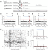
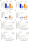
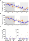
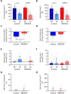
The pivotal role of the periaqueductal grey (PAG) in fear learning is reinforced by the identification of neurons in male rat ventrolateral PAG (vlPAG) that encode fear memory through signalling the onset and offset of an auditory-conditioned stimulus during presentation of the unreinforced conditioned tone (CS+) during retrieval. Some units only display CS+ onset or offset responses, and the two signals differ in extinction sensitivity, suggesting that they are independent of each other. In addition, understanding cerebellar contributions to survival circuits is advanced by the discovery that (i) reversible inactivation of the medial cerebellar nucleus (MCN) during fear consolidation leads in subsequent retrieval to (a) disruption of the temporal precision of vlPAG offset, but not onset responses to CS+, and (b) an increase in duration of freezing behaviour. And (ii) chemogenetic manipulation of the MCN-vlPAG projection during fear acquisition (a) reduces the occurrence of fear-related ultrasonic vocalisations, and (b) during subsequent retrieval, slows the extinction rate of fear-related freezing. These findings show that the cerebellum is part of the survival network that regulates fear memory processes at multiple timescales and in multiple ways, raising the possibility that dysfunctional interactions in the cerebellar-survival network may underlie fear-related disorders and comorbidities.
Keywords: PAG; cerebellum; fear; neuroscience; rat; survival circuits; timing.
Plain language summary
Anxiety disorders are a cluster of mental health conditions characterised by persistent and excessive amounts of fear and worry. They affect millions of people worldwide, but treatments can sometimes be ineffective and have unwanted side effects. Understanding which brain regions are involved in fear and anxiety-related behaviours, and how those areas are connected, is the first step towards designing more effective treatments. A region known as the periaqueductal grey (or PAG) sits at the centre of the brain’s fear and anxiety network, regulating pain, encoding fear memories and responding to threats and stressors. It also controls survival behaviours such as the ‘freeze’ response, when an animal is frightened. A more recent addition to the fear and anxiety network is the cerebellum, which sits at the base of the brain. Two-way connections between this region and the PAG have been well described, but how the cerebellum might influence fear and anxiety-related behaviours remains unclear. To explore this role, Lawrenson, Paci et al. investigated whether the cerebellum modulates brain activity within the PAG and if so, how this relates to fear behaviours. Rats had electrodes implanted in their brains to record the activity of nerve cells within the PAG. A common fear-conditioning task was then used to elicit ‘freeze’ responses: a sound was paired with mild foot shocks until the animals learned to fear the auditory signal. In the rats, a subset of neurons within the PAG responded to the tone, consistent with those cells encoding a fear memory. But when a drug blocked the cerebellum’s output during fear conditioning, the timing of the PAG response was less precise and the rats’ freeze response lasted longer. Lawrenson, Paci et al. concluded that the cerebellum, through its interactions with the brain’s fear and anxiety network, might be responsible for coordinating the most appropriate behavioural response to fear, and how long ‘freezing’ lasts. In summary, these findings show that the cerebellum is a part of the brain’s survival network which regulates fear-memory processes. It raises the possibility that disruption of the cerebellum might underlie anxiety and other fear-related disorders, thereby providing a new target for future therapies.
© 2022, Lawrenson et al.
Conflict of interest statement
CL, EP, JP, RD, BL, RA No competing interests declared
Figures





References
-
- An X, Bandler R, Ongür D, Price JL. Prefrontal cortical projections to longitudinal columns in the midbrain periaqueductal gray in macaque monkeys. The Journal of Comparative Neurology. 1998;401:455–479. - PubMed
-
- Asdourian D, Frerichs K. Some effects of cerebellar stimulation. Psychonomic Science. 1970;18:261–262. doi: 10.3758/BF03331817. - DOI
Publication types
MeSH terms
Grants and funding
LinkOut - more resources
Full Text Sources
Research Materials

