Review
. 2022 Jul:245:55-81.
doi: 10.1016/j.trsl.2022.03.004. Epub 2022 Mar 12.

Cell type-specific mechanistic target of rapamycin-dependent distortion of autophagy pathways in lupus nephritis

Affiliations
Review

Cell type-specific mechanistic target of rapamycin-dependent distortion of autophagy pathways in lupus nephritis

Tiffany Caza et al. Transl Res. 2022 Jul.

Abstract

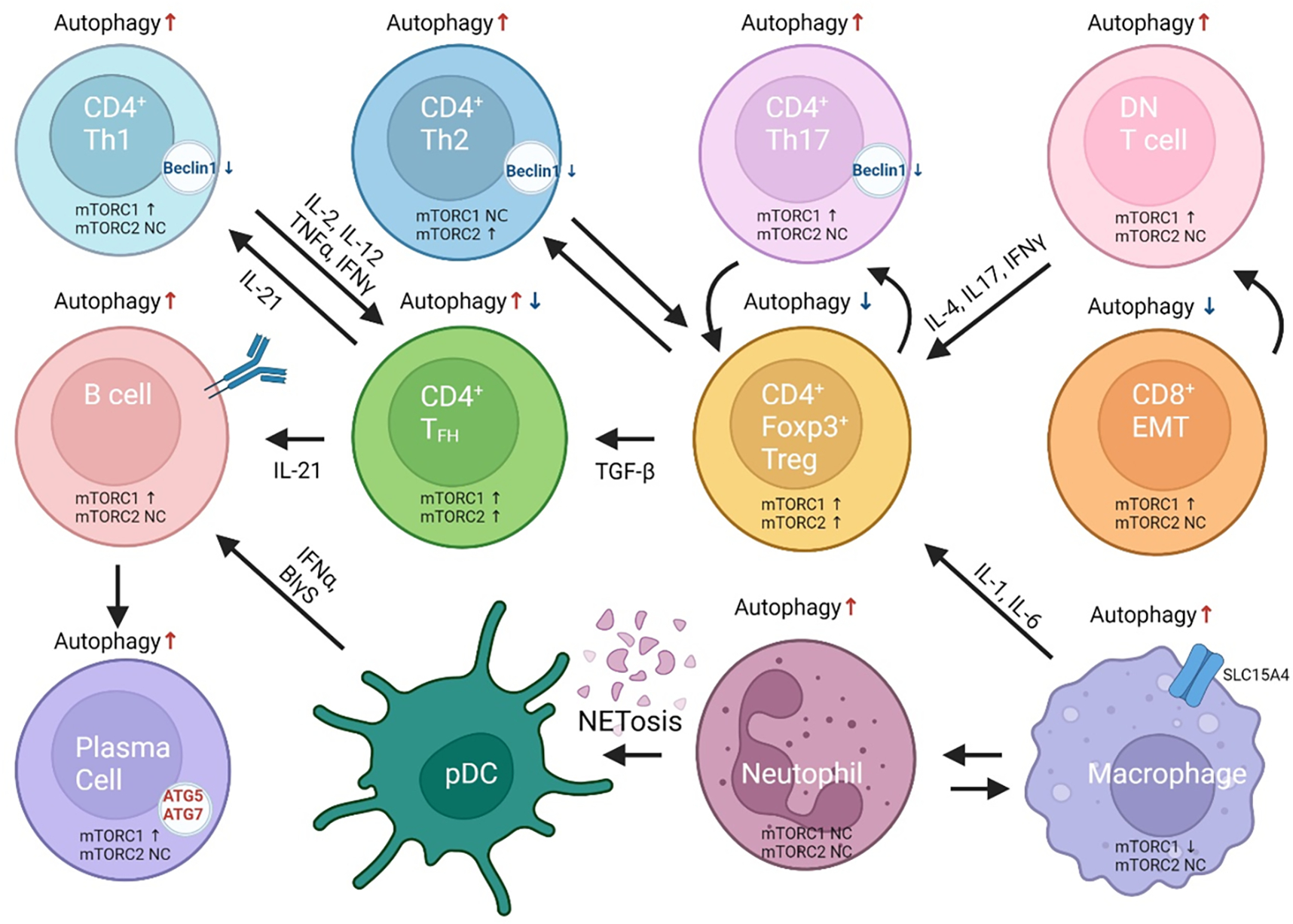
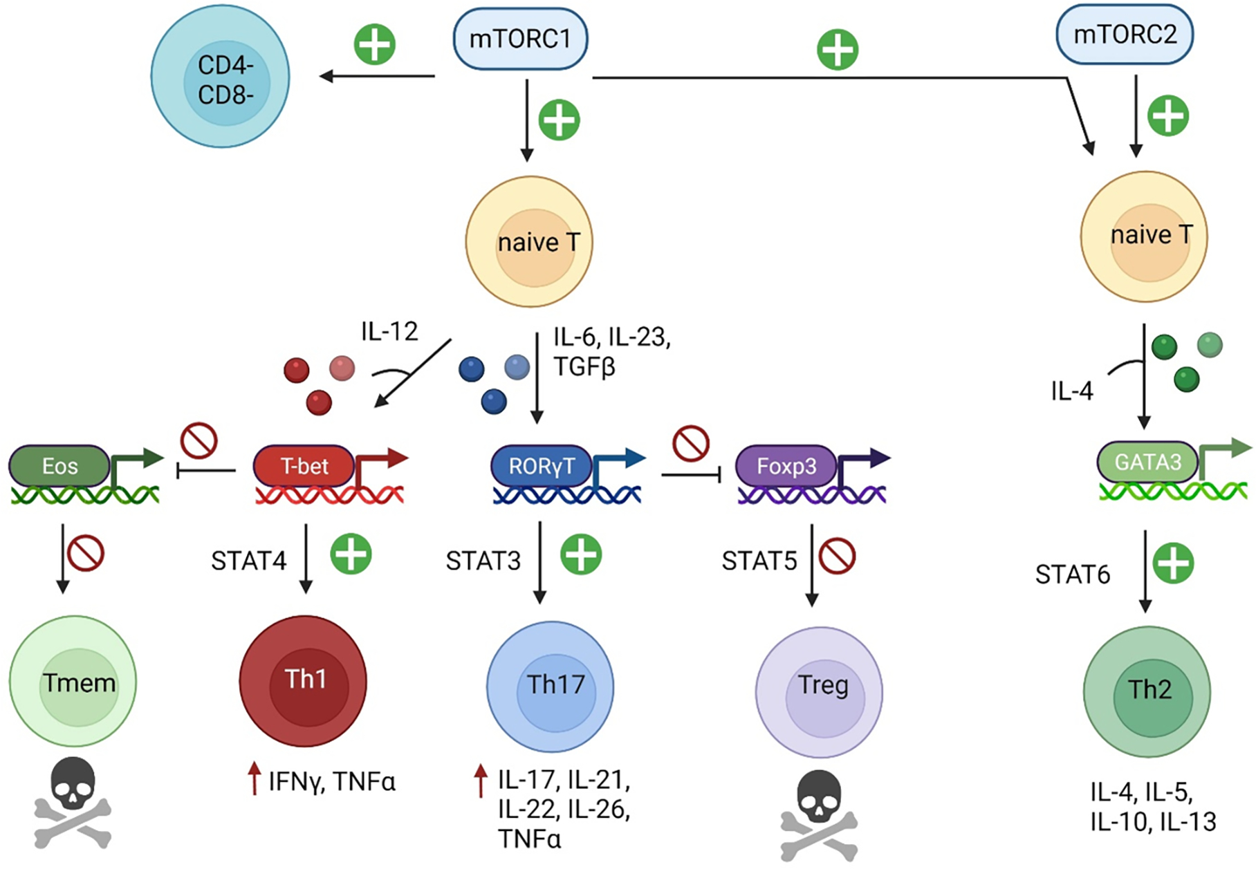
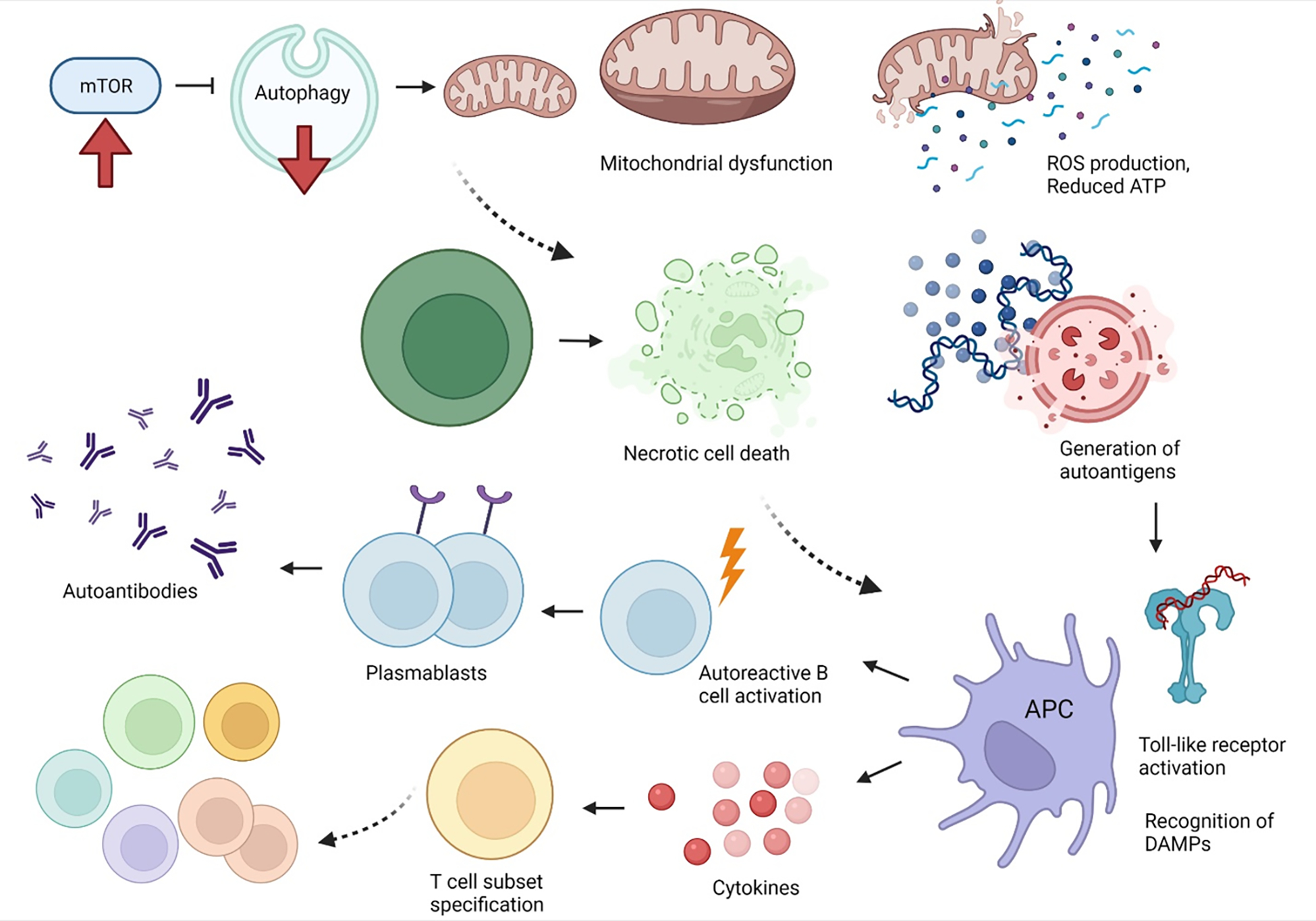
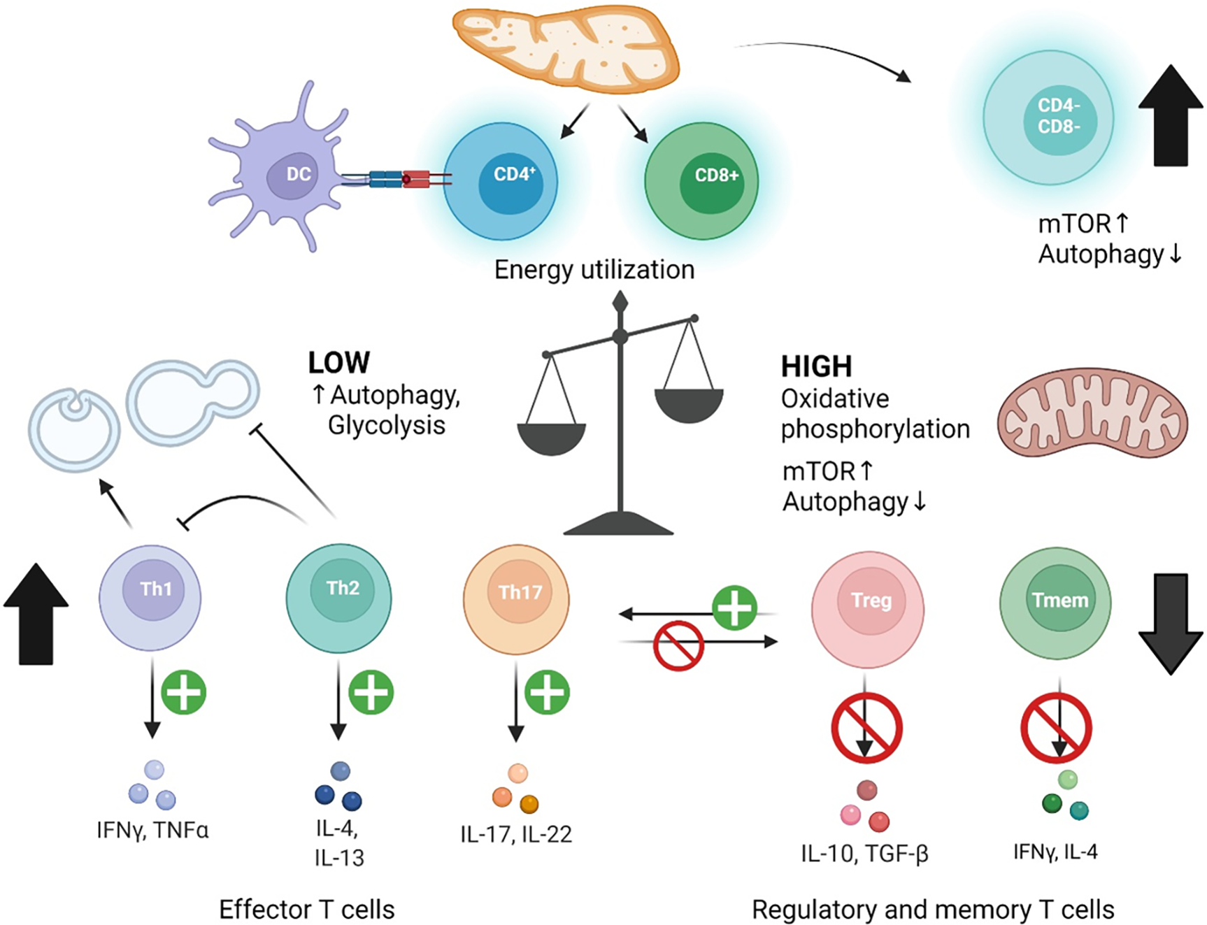
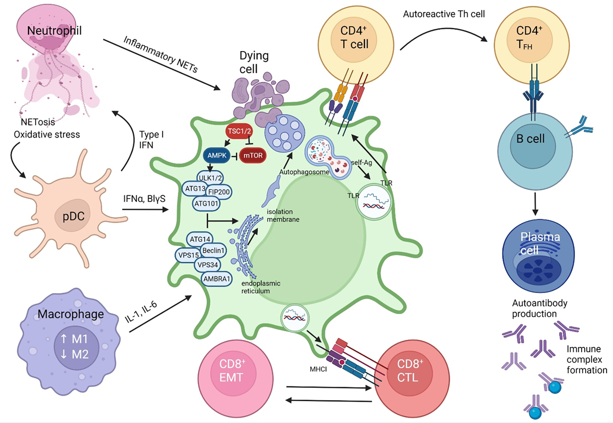
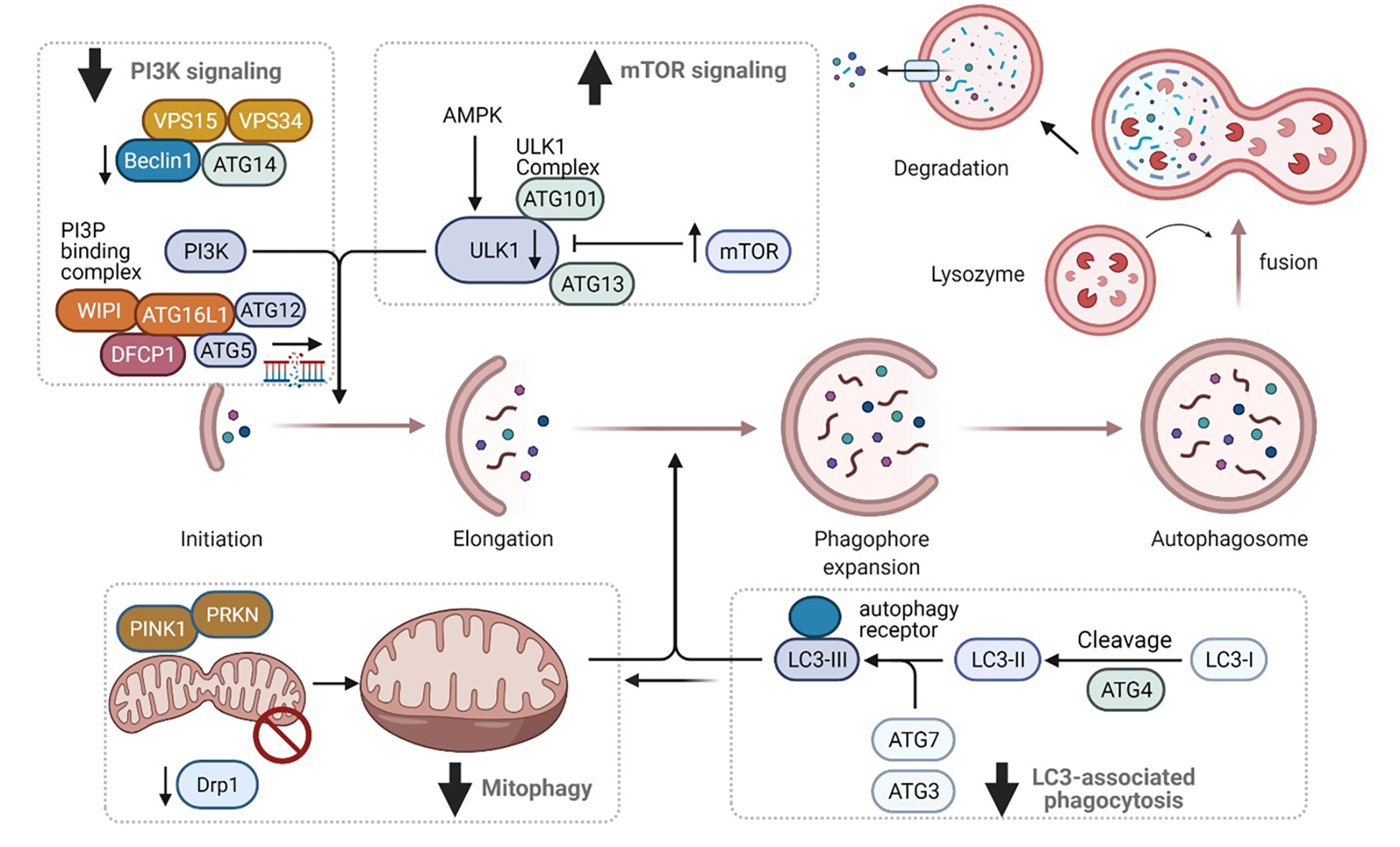
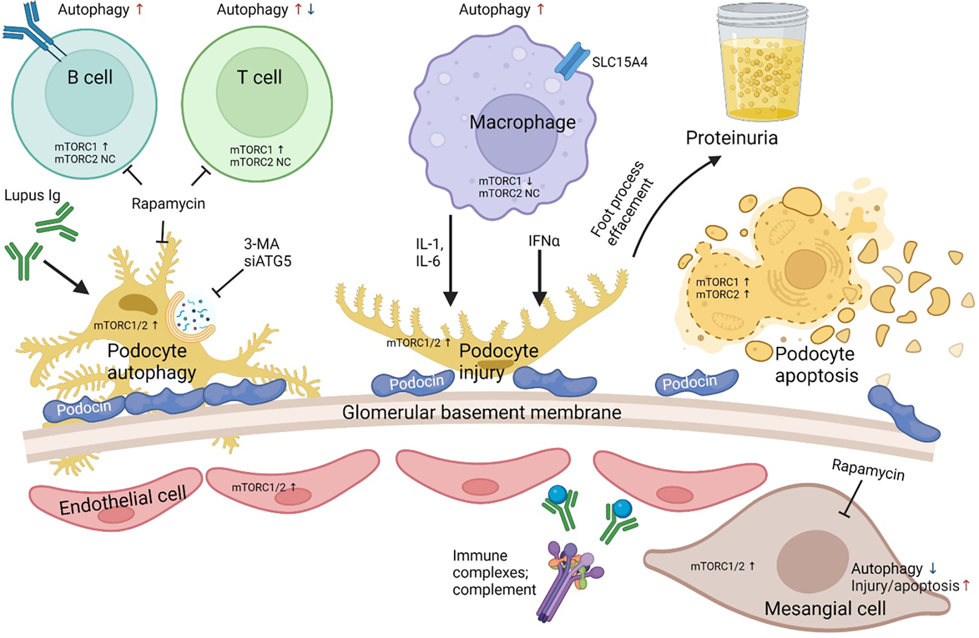
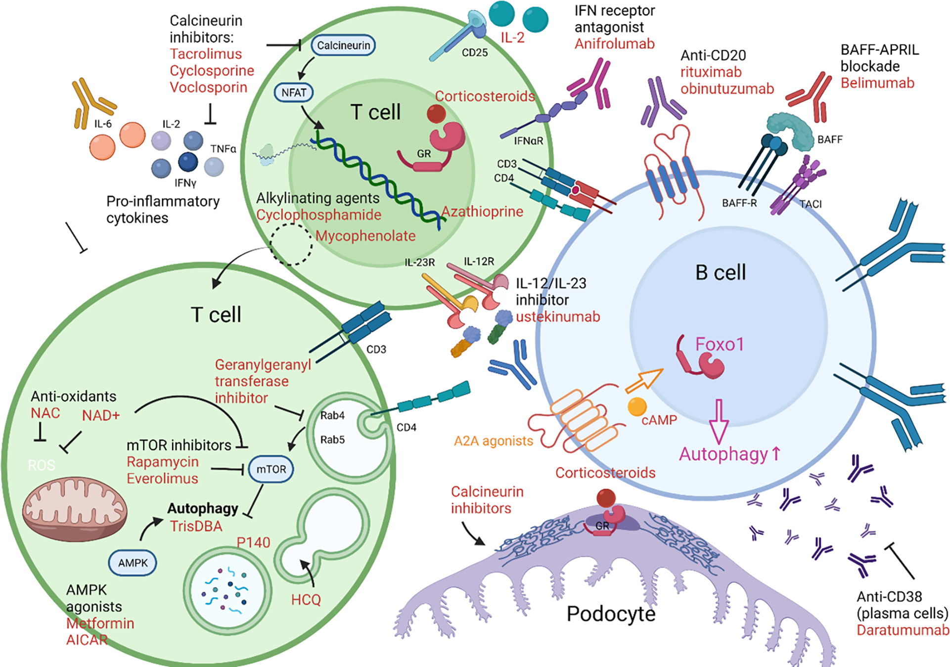
Pro-inflammatory immune system development, metabolomic defects, and deregulation of autophagy play interconnected roles in driving the pathogenesis of systemic lupus erythematosus (SLE). Lupus nephritis (LN) is a leading cause of morbidity and mortality in SLE. While the causes of SLE have not been clearly delineated, skewing of T and B cell differentiation, activation of antigen-presenting cells, production of antinuclear autoantibodies and pro-inflammatory cytokines are known to contribute to disease development. Underlying this process are defects in autophagy and mitophagy that cause the accumulation of oxidative stress-generating mitochondria which promote necrotic cell death. Autophagy is generally inhibited by the activation of the mammalian target of rapamycin (mTOR), a large protein kinase that underlies abnormal immune cell lineage specification in SLE. Importantly, several autophagy-regulating genes, including ATG5 and ATG7, as well as mitophagy-regulating HRES-1/Rab4A have been linked to lupus susceptibility and molecular pathogenesis. Moreover, genetically-driven mTOR activation has been associated with fulminant lupus nephritis. mTOR activation and diminished autophagy promote the expansion of pro-inflammatory Th17, Tfh and CD3+CD4-CD8- double-negative (DN) T cells at the expense of CD8+ effector memory T cells and CD4+ regulatory T cells (Tregs). mTOR activation and aberrant autophagy also involve renal podocytes, mesangial cells, endothelial cells, and tubular epithelial cells that may compromise end-organ resistance in LN. Activation of mTOR complexes 1 (mTORC1) and 2 (mTORC2) has been identified as biomarkers of disease activation and predictors of disease flares and prognosis in SLE patients with and without LN. This review highlights recent advances in molecular pathogenesis of LN with a focus on immuno-metabolic checkpoints of autophagy and their roles in pathogenesis, prognosis and selection of targets for treatment in SLE.


Conflict of interest statement

The authors have read the journal’s policy on disclosure of potential conflicts of interest and declare no conflicts of interest. All authors have read the journal’s authorship agreement and that the manuscript has been reviewed by and approved by all named authors.

Figures

Figure 1.
Figure 1.. Coordinate skewing of mTOR activation and autophagy in cells of the adaptive and innate immune systems underlie pro-inflammatory lineage specification in SLE.
mTOR activation is considered as a central mediator of disease pathogenesis. There is a direct and well established relationship between mTOR activity and autophagy, with increased activity resulting in diminished autophagy. Different T-cell lineages, however, have discordant skewing in autophagy. Naive CD4+ T cells have increased autophagy. CD4+ Th17 cells have decreased autophagy and increased mTORc1 activity. CD4+FoxP3+Treg cells have diminished autophagy with increased activation of both complexes, mTORC1 and mTORC2. CD4+Tfh cells have mixed results with increased or decreased autophagy but mTOR complexes are both increased. CD8+ cytotoxic T cells have increased autophagy resulting in increased cell death. Activity of their mTOR complexes is not fully known. CD8 effector memory T (EMT) cells have reduced autophagy and activation of mTORC1 which are responsive to rapamycin treatment in vivo and in vitro. B cells and plasma cells have increased autophagy required for B cell differentiation, with spurious elevation of MTORC1.
Figure 2.
Figure 2.. mTOR activation mediates pro-inflammatory T-cell lineage specification in SLE.
Activation of mTORC1 in naïve lupus T cells results in CD4+ T cell specification to the Th1 and Th17 lineages, through activation of lineage specific transcription factors in the presence of polarizing cytokines. This results in activation of JAK-STAT signaling and production of cytokines and other inflammatory mediators. T-bet expression during Th1 specification inhibits eomesodermin, impairing differentiation to the memory T cell lineage. Additionally, RORγT expression for Th17 specification results in reduced expression of Foxp3, inhibiting Treg development. mTORC2 activation promotes Th2 specification through the transcription factor GATA-3 in the presence of IL-4.
Figure 3.
Figure 3.. Overview of changes in autophagy during lupus pathogenesis.
mTOR activation with subsequent inhibition of autophagy is central to lupus pathogenesis. Deficient autophagy, including autophagic turnover of mitochondria (mitophagy), results in mitochondrial dysfunction. Mitochondrial dysfunction is characterized by increased mitochondrial mass, mitochondrial hyperpolarization, increased production of reactive oxygen species, and reduced production of ATP. Upon sustained T-cell activation, mitochondrial dysfunction predisposes to necrotic cell death, rather than apoptosis. Released nucleic acids and other intracellular materials can serve as damage-associated molecular patterns (DAMPs) or autoantigens, inducing toll-like receptor activation within antigen-presenting cells. Activation of antigen presenting cells results in activation of autoreactive B cells which can differentiate into plasmablasts to produce autoantibodies. Additionally, cytokine production can preserve CD4−CD8− T cells and skew T-cell subset specification towards inflammatory Th1, Th2, Th17 cells, rather than Tregs or memory T cells.
Figure 4.
Figure 4.
Role of autophagy in pro-inflammatory immune cell specification in SLE. Immune cell subsets have differential requirements for autophagy in activation and maintenance, of which is primarily regulated by mTOR activation and is dependent on energy metabolism. CD4CD8 T cells of which escape thymic selection show increased mTOR activity and deficient autophagy. CD4+ Tregs and CD8+ memory T cells also have increased mTOR activity, deficient autophagy, and are depleted in SLE. Tregs are additionally depleted through increased differentiation into Th17 cells. Dendritic cells have increased autophagy that increases antigen presentation to naïve CD4+ T cells. Th subsets with low energy requirements utilize glycolysis and turnover cellular components by autophagy. Th1 and Th17 cells show activated autophagy. Th2 cells produce IL-4 and IL-13 which in turn inhibits autophagy and IFNγ production by Th1 cells.
Figure 5.
Figure 5.. Defective canonical autophagy underlies pathogenic cell-to-cell communication in SLE.
In a susceptible host, genetic material from apoptotic cells primes an autoreactive immune response. NETosis and oxidative activity increases in neutrophils. The NET DNA detection by pDC causes an increase in type 1 IFN signalling, which is a hallmark of SLE disease (135, 136).. IFNα signalling, along with the increased secretion of pro-inflammatory cytokines IL-6, IL-17, IL-18, IL-23 and type 1 IFN stimulates native dendritic cells. Homeostatic balance between macrophages shift to favor the M1 differentiation with a resultant pro-inflammatory cascade. Endocytosis of a dying cell, in the susceptible host with defective components of autophagy will result in the non-fusion of the phagosome with the lysosome. The pre-initiation complex of autophagosome formation is comprised of ULK1/2, ATG13, FIP200 and ATG101. Interaction with the complex of VPS15, ATG14, BECN1, VPS34 and AMBRA1 generates the isolation membrane from the endoplasmic reticulum or mitochondria which is required for phagosome formation (90). Defects in BECN1, CYBB/NOX2, RUBCN, ATG7, ATG5 are associated with reduced autophagosome formation and defective clearance of dying cells that trigger inflammation in SLE and lupus nephritis (90, 122, 123, 125). AMPK activates while mTOR inhibits autophagy via phosphorylation of ULK1. In turn, TSC1 and TSC2 restrain mTOR activation. The inability to complete autophagy into the final autolysosome step causes the release of self-antigens which are then presented by antigen presenting cells to CD4+ helper T cells and CD8+ cytotoxic T cells via MHC class II and MHC class I, respectively. The result is the stimulation of the adaptive immune system towards autoreactive helper T-cells and cytotoxic T-cells which lead to tissue damage. Primed CD4+ T cells form and immune synapse with and release cytokines, IL-4 and IL-21 to activate B cells which differentiate to immunoglobulin-secreting plasma cells. FN: Interferon, NET: Neutrophil extracellular traps, ER: endoplasmic reticulum, DNA: Deoxyribonucleic acid, pDC: plasmacytoid dentritic cell, CD28 & B7: costimulatory signals, TLR: toll like receptor, BCR: B-cell receptor.
Figure 6.
Figure 6.. Molecular checkpoints of distorted autophagy pathways in lupus nephritis
The process and main regulatory mechanisms of autophagy. The process of autophagy starts with cytoplasmic material being engulfed by double membranes with the formation of cup-like structure called the phagophore to the conversion into double membrane vesicles called the autophagosomes. The mTOR pathway and the AMP- activated Kinase (AMPK) are key determinants of autophagy activation. The process if finely regulated by autophagy-related proteins (ATG) that assemble into several complexes : Unc-51-Like Kinase (ULK1), PI3K nucleation complex and the phosphatidinol 3 phosphate binding complex. Together, they direct the distribution of the autophagy machinery allowing the formation of the autophagosome. LC3 is cleaved into LC3-I by ATG4 which is then conjugated to phosphatidylethanolamine to form LC3-II. This complex gets incorporated into autophagosome and binds with receptors harboring LC3 containing motifs. Both macroautophagy and LC3-associated autophagy can impact mitophagy, of which is largely deficient in SLE, but dependent on the energy requirements of effector cell types.
Figure 7.
Figure 7.. mTOR-dependent changes in autophagy involve renal and infiltrating-inflammatory cells in patients with lupus nephritis.
Inhibition of ATG5 by small interfering RNA and 3-MA inhibits autophagy in podocytes resulting in an aggravation of podocyte apoptosis and injury. Sera obtained from lupus nephritis patients demonstrated an increase in podocin by IFNα and an increase in podocyte injury and apoptosis by IgG. The combined cellular injury and podocyte foot process effacement leads to increase in proteinuria. Rapamycin which is a potent inhibitor of mTOR induces autophagy in such patients and reconstitute the podocyte foot processes and diminishes podocyte apoptosis leading to a decrease in albumin flux and proteinuria. mTORC1, mTORC2 and autophagy is skewed in a cell type-specific manner within the innate and adaptive immune system of SLE patients (58, 59, 178, 179).
Figure 8.
Figure 8.. Therapeutic targeting of autophagy in lupus nephritis.
Targeting metabolism expands our repertoire of potential therapeutics in SLE. Traditional therapies have targeted DNA replication (cyclophosphamide, mycophenolate mofetil, azathioprine) or inhibiting T-cell activation through inhibiting calcineurin which prevents production of IL-2 and other cytokines (tacrolimus, cyclosporine, voclosporin), or through activating glucocorticoid receptors (corticosteroids, such as prednisone or methylprednisolone). Calcineurin inhibitors and corticosteroids can also function to stabilize the podocyte cytoskeleton. Aberrations in T-cell metabolism can be targeted through mTOR inhibition (everolimus, sirolimus/rapamycin), which restores autophagy. Anti-oxidants can inhibit mTOR, including NAC or NAD+. Autophagy can be activated through treatment with TrisDBA or agonists of AMPK (metformin, AICAR). It is also affected through altering endosomal acidification with anti-malarial drugs (hydroxychloroquine). Endosomal function can also be targeted by inhibiting RabGTPases through geranylgeranyl transferase inhibitors. Cytokines and cytokine receptors can be targeted for therapy, including the p40 domain of IL-2 and IL-23 (ustekinumab), interferon receptor (anifrolumab), and low-dose IL-2 therapy. The JAK inhibitor, baricitinib, also reduces production of pro-inflammatory cytokines through blockade of JAK-STAT signaling. Autoantibody production can be reduced through depletion of B cells by monoclonal antibodies directed against CD20 (rituximab, obinutuzumab) or depletion of plasma cells/plasmablasts with anti-CD38 monoclonal antibodies (daratumumab). Alternatively, B cell activation can be inhibited through the BAFF-APRIL axis (belimumab).

Similar articles

Cited by

References

    1. Alarcón GS, Roseman J, Bartolucci AA, Friedman AW, Moulds JM, Goel N, et al. Systemic lupus erythematosus in three ethnic groups: II. Features predictive of disease activity early in its course. LUMINA Study Group. Lupus in minority populations, nature versus nurture. Arthritis Rheum. 1998;41(7):1173–80. Epub 1998/07/15. doi: 10.1002/1529-0131(199807)41:7<1173::Aid-art5>3.0.Co;2-a. - DOI - PubMed
    1. Madhok R Systemic lupus erythematosus: lupus nephritis. BMJ Clin Evid. 2015;2015. Epub 2015/12/20. - PMC - PubMed
    1. Mok CC. Biomarkers for lupus nephritis: a critical appraisal. J Biomed Biotechnol. 2010;2010:638413. Epub 2010/04/24. doi: 10.1155/2010/638413. - DOI - PMC - PubMed
    1. Aragón CC, Tafúr RA, Suárez-Avellaneda A, Martínez MT, Salas AL, Tobón GJ. Urinary biomarkers in lupus nephritis. J Transl Autoimmun. 2020;3:100042. Epub 2020/08/04. doi: 10.1016/j.jtauto.2020.100042. - DOI - PMC - PubMed
    1. Mahmoud SS, Bazaraa HM, Lotfy HM, Abd-El-Aziz DM. Renal involvement in childhood-onset systemic lupus erythematosus in Egypt. Rheumatol Int. 2012;32(1):47–51. Epub 2010/07/27. doi: 10.1007/s00296-010-1554-7. - DOI - PubMed

Publication types