Review
. 2022 Nov 1;12(11):a041183.
doi: 10.1101/cshperspect.a041183.

Endothelial Cell Fate Determination: A Top Notch Job in Vascular Decision-Making

Affiliations
Review

Endothelial Cell Fate Determination: A Top Notch Job in Vascular Decision-Making

L A Naiche et al. Cold Spring Harb Perspect Med. .

Abstract

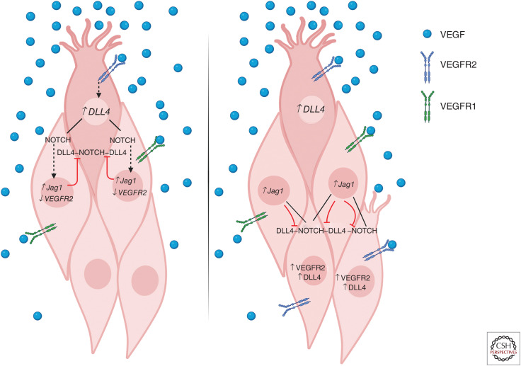
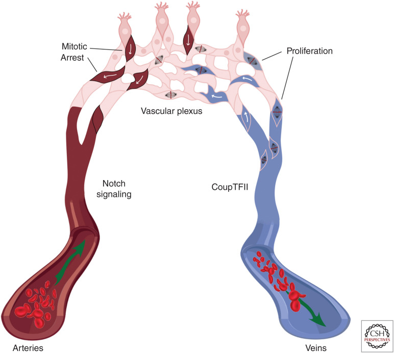
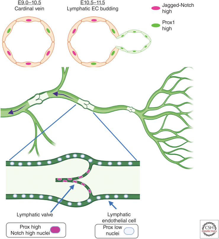
As vascular networks form, endothelial cells (ECs) undergo cell fate decisions that determine whether they become tip or stalk cells of the developing vascular plexus or mature into arterial, venous, or lymphatic endothelium. EC fate decisions are coordinated with neighboring cells to initiate sprouting, maintain endothelial barrier, or ensure appropriate specialization of vessels. We describe mechanisms that control EC fate at specific steps in these processes, with an emphasis on the role of the Notch signaling pathway. Specific EC fate determination steps that are highlighted are tip/stalk selection during sprouting angiogenesis, venous-arterial specification, arteriogenesis, lymphatic vessel specification, and lymphatic valve formation.


Figures

Figure 1.
Figure 1.
Tip cell/stalk cell determination and shuffling in sprouting angiogenesis. (Left) Upon initiation of tip cell differentiation in response to vascular endothelial growth factor (VEGF)-VEGFR2 signaling, expression of Notch ligand DLL4 is increased. DLL4 signaling via Notch receptors in adjacent cells represses DLL4 and VEGFR2 expression and up-regulates VEGFR1 and Jagged1 expression, enforcing the stalk cell identity of neighboring cells and preventing unregulated tip cell sprouting. Jagged1 expression in neighboring stalk cells inhibits DLL4-Notch signaling in the tip cell, further stabilizing tip/stalk identity in the local area. (Right) Distal to the leading tip cell, stalk cells that receive lower levels of DLL4-Notch signaling and highly express VEGFR2 retain the ability to respond to VEGF stimulation and adopt tip cell identity. Upon VEGF stimulation, these cells may compete with the previously specified tip cell for position at the leading edge of the vascular front.
Figure 2.
Figure 2.
Arterial/venous differentiation from the vascular plexus. Arterial endothelial cells (ECs) (left) express Notch proteins, while venous ECs (right) have low Notch expression and high CoupTFII expression. CoupTFII drives endothelial proliferation in venous endothelium, extending the vein and contributing cells to the growing vascular plexus (top). Stalk cells in the vascular plexus experience high levels of Notch signaling, which causes cell-cycle arrest. High Notch signaling and cell-cycle arrest leads the cell to assume arterial fate, migrate against the direction of blood flow, and incorporate into the artery.
Figure 3.
Figure 3.
Acquisition of lymphatic endothelial and lymphatic valve cell fates. (Top) During murine embryonic development, Prox1-expressing cells form in the cardinal vein (right side on schematized vein) and bud to form a sprout that initiates lymphatic development. Notch1 and Jagged1-expressing cells are located at the opposite side of the vein from Prox1-expressing cells and loss-of-function experiments demonstrate that Notch suppresses Prox1 expression, assuring proper lymphatic development. (Bottom) Lymphatic vasculature consists of capillaries that collect fluid, which is directed through lymphatic ducts containing valves that assure unidirectional flow. During initial development of lymphatic valves, lymphatic endothelial cells (ECs) express high levels of Prox1 and have high Notch signaling, and loss of Notch1 or canonical Notch signaling prevents proper lymphatic valve development.

References

    1. Arima S, Nishiyama K, Ko T, Arima Y, Hakozaki Y, Sugihara K, Koseki H, Uchijima Y, Kurihara Y, Kurihara H. 2011. Angiogenic morphogenesis driven by dynamic and heterogeneous collective endothelial cell movement. Development 138: 4763–4776. 10.1242/dev.068023 - DOI - PubMed
    1. Armulik A, Genové G, Mäe M, Nisancioglu MH, Wallgard E, Niaudet C, He L, Norlin J, Lindblom P, Strittmatter K, et al. 2010. Pericytes regulate the blood–brain barrier. Nature 468: 557–561. 10.1038/nature09522 - DOI - PubMed
    1. Bazigou E, Xie S, Chen C, Weston A, Miura N, Sorokin L, Adams R, Muro AF, Sheppard D, Makinen T. 2009. Integrin-α9 is required for fibronectin matrix assembly during lymphatic valve morphogenesis. Dev Cell 17: 175–186. 10.1016/j.devcel.2009.06.017 - DOI - PMC - PubMed
    1. Benedito R, Roca C, Sörensen I, Adams S, Gossler A, Fruttiger M, Adams RH. 2009. The Notch ligands Dll4 and Jagged1 have opposing effects on angiogenesis. Cell 137: 1124–1135. 10.1016/j.cell.2009.03.025 - DOI - PubMed
    1. Benedito R, Rocha SF, Woeste M, Zamykal M, Radtke F, Casanovas O, Duarte A, Pytowski B, Adams RH. 2012. Notch-dependent VEGFR3 upregulation allows angiogenesis without VEGF-VEGFR2 signalling. Nature 484: 110–114. 10.1038/nature10908 - DOI - PubMed

Publication types

Substances

LinkOut - more resources