Case Reports
. 2022 Mar 14;15(3):e248505.
doi: 10.1136/bcr-2021-248505.

A Low rectal GIST

Affiliations
Case Reports

A Low rectal GIST

Tara M Connelly et al. BMJ Case Rep. .

Abstract

A man in his 70s presented with narrowing of his stool calibre and a palpable mass on rectal examination. Colonoscopy revealed a submucosal bulge without a mucosal lesion. CT and MRI demonstrated an 8×5.4×7 cm mass in lower rectum. Biopsy of the mass confirmed a rectal gastrointestinal stromal tumour (GIST). It was moderately sensitive to neoadjuvant imatinib and radiotherapy, which slightly downsized the tumour. He required abdominal perineal resection with curative intent. Uncommonly found in the rectum, when located here, GISTs are typically aggressive and are a rare cause of stool calibre narrowing. Rectal GISTs should be considered in patients presenting with this symptom.

Keywords: gastrointestinal surgery; gastrointestinal system; malignant disease and immunosuppression; radiology (diagnostics).


Conflict of interest statement

Competing interests: None declared.

Figures

Figure 1
Figure 1
Colonoscopic view of the GIST protruding into the rectum. An adenoma is visible proximally (arrow).
Figure 2
Figure 2
Coronal and axial CT images depicting the rectal GIST (arrow). GIST, gastrointestinal stromal tumour.
Figure 3
Figure 3
Axial MRI depicting the well circumscribed GIST compressing the lesion. GIST, gastrointestinal stromal tumour.
Figure 4
Figure 4
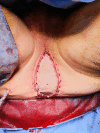
Perineal views of en bloc specimen extraction (A) Patient positioning in the prone Kraske position with well-padded pressure points; (B) preincision elliptical marking around the anus (C) transperineal specimen extraction and (D) defect after resection of the coccyx (marked with*), levators, lesion and anus.
Figure 5
Figure 5
Views of abdomen: (A) VRAM flap harvesting with end colostomy site located to the patient’s left and laterally (marked with *); (B) VRAM donor site closure with mesh.
Figure 6
Figure 6
VRAM flap placement in perineal defect.
Figure 7
Figure 7
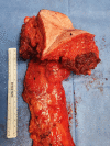
Abdominal perineal resection specimen.
Figure 8
Figure 8
Staining of rectal GIST (A) H&E×4, (B) H&E×20, (C) CD117/KIT (D) DOG1.

References

    1. Kelley KA, Byrne R, Lu KC,. Gastrointestinal stromal tumors of the distal gastrointestinal tract. Clin Colon Rectal Surg 2018;31:295–300. 10.1055/s-0038-1642053 - DOI - PMC - PubMed
    1. Network NCC, 2021. Available: https://www.nccn.org/professionals/physician_gls/pdf/gist.pdf [Accessed 1 Dec 2021].
    1. Ishikawa T, Kanda T, Kameyama H, et al. . Neoadjuvant therapy for gastrointestinal stromal tumor. Transl Gastroenterol Hepatol 2018:3:3. - PMC - PubMed
    1. Kameyama H, Kanda T, Tajima Y, et al. . Management of rectal gastrointestinal stromal tumor. Transl Gastroenterol Hepatol 2018;3:8. 10.21037/tgh.2018.01.08 - DOI - PMC - PubMed
    1. Jiang Z-X, Zhang S-J, Peng W-J, et al. . Rectal gastrointestinal stromal tumors: imaging features with clinical and pathological correlation. World J Gastroenterol 2013;19:3108–16. 10.3748/wjg.v19.i20.3108 - DOI - PMC - PubMed

Publication types

MeSH terms