. 2022 Mar 14;14(5):2418-2431.
doi: 10.18632/aging.203951. Epub 2022 Mar 14.

miRNA-29a inhibits atherosclerotic plaque formation by mediating macrophage autophagy via PI3K/AKT/mTOR pathway

Affiliations

miRNA-29a inhibits atherosclerotic plaque formation by mediating macrophage autophagy via PI3K/AKT/mTOR pathway

Weihua Shao et al. Aging (Albany NY). .

Abstract

Background: miR-29a plays a vital role in AS, but the relationship between the miR-29a-targeted PI3K signaling pathway and AS remains unclear. Therefore, this study was carried out.

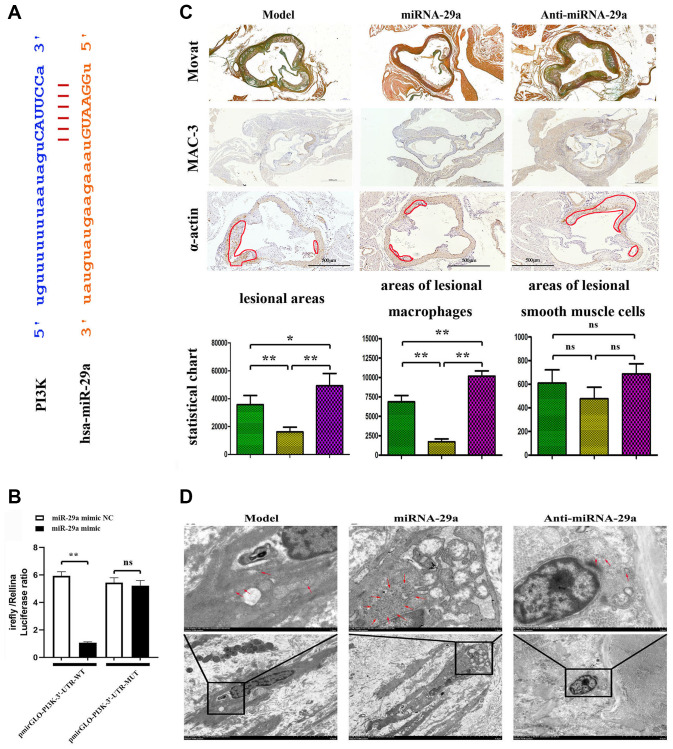
Methods: Gene expression profiles from the GEO database containing AS samples were analyzed. ApoE-/- mice and RAW264.7 cells were treated with miR-29a negative control (NC), miR-29a mimic and miR-29a inhibitor to establish the AS model. Then MOVAT staining, TEM, Western blotting, and immunofluorescence staining were adopted for testing target proteins.

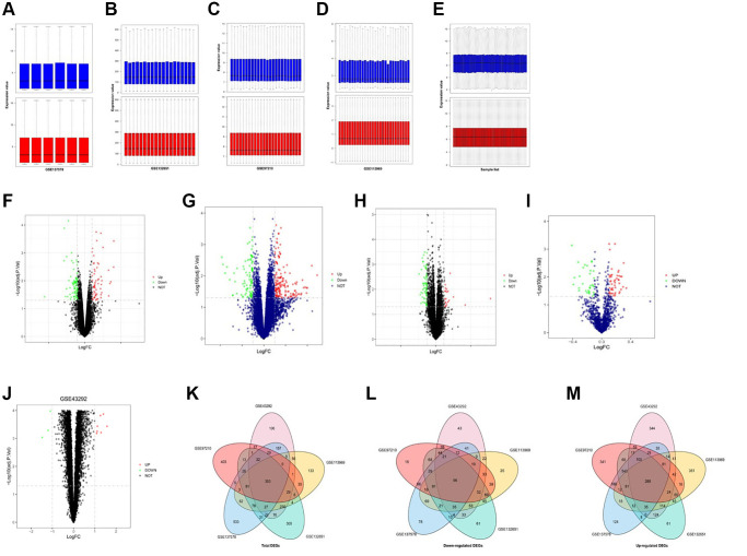
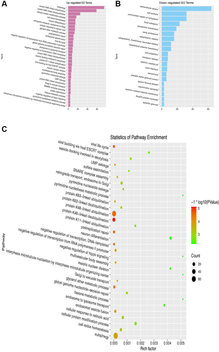
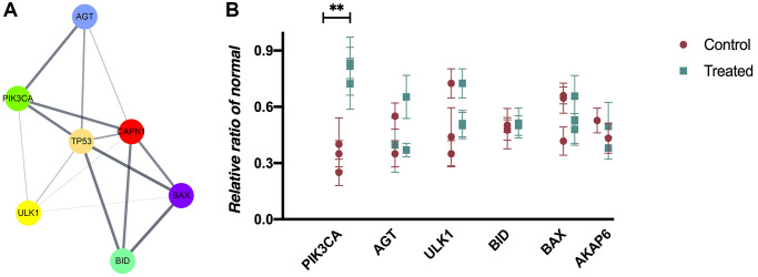
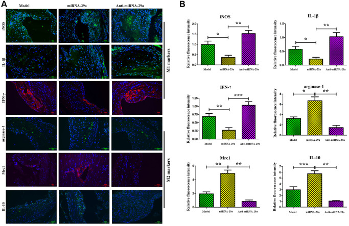
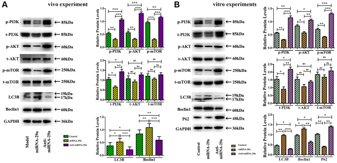
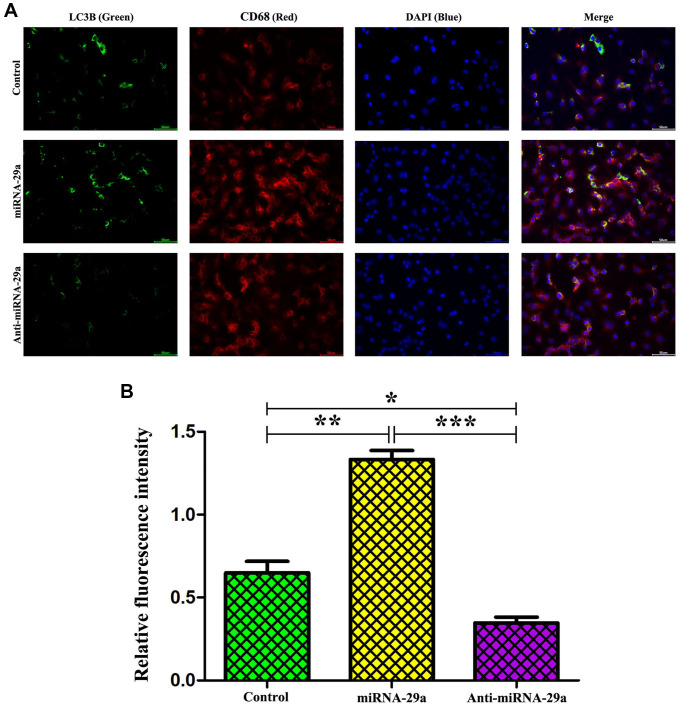
Results: DEGs were identified from GSE137578, GSE132651, GSE113969, GSE43292, and GSE97210 datasets. It was found that there were targeted binding sites between miR-29a and PIK3CA. Besides, GO and KEGG analysis demonstrated that autophagy was an enriched pathway in AS. Later, PPI network was depicted, and hub genes were then determined. The results revealed that miR-29a suppressed the areas of plaques and lesional macrophages, but had no impact on VSMCs. TEM results showed the organelles pyknosis of lesional macrophages damaged morphological changes. Furthermore, miR-29a amplified the M2-like macrophages but suppressed the polarization of M1-like macrophages in atherosclerotic plaques. According to mouse and RAW 264.7 cell experiments, miR-29a significantly inhibited the protein expressions of PI3K, p-PI3K, p-AKT, and p-mTOR, which were consistent with the increased expressions of autophagy-related proteins, Beclin 1 and LC3II. However, the miR-29a suppression exhibited the contrary results.

Conclusion: MiR-29a elevation induces the increase of autophagy by down-regulating the PI3K/AKT/mTOR pathway in the progression of AS, indicating that miR-29a is a novel therapeutic strategy for AS.

Keywords: PI3K/AKT/mTOR; atherosclerosis; atherosclerotic; macrophage; miR-29a; plaque.


Conflict of interest statement

CONFLICTS OF INTEREST: The authors declare that they have no known competing financial interests or personal relationships that could have influenced the work reported in this paper.

Figures

Figure 1
Figure 1
Identification and integration of DEGs in AS. (AE) Pre-standardization and post-standardization gene expressions of GSE137578, GSE132651, GSE113969, GSE43292 and GSE97210 are demonstrated. (EH) DEGs of each dataset are assessed by criteria of P < 0.05 and |log2FC|≥2. (FJ) Volcano maps of the top 100 DEGs of GSE137578, GSE132651, GSE113969, GSE97210 and GSE43292. (KM) The total upregulated and downregulated DEGs of GSE137578, GSE132651, GSE113969, GSE43292, and GSE97210 are overlapped and depicted by the Venn diagram analysis.
Figure 2
Figure 2
GO and KEGG pathway enrichment analyses of DEGs. (A, B) GO enrichment analysis of overlapping DEGs. (C) KEGG pathway enrichment analysis of overlapping DEGs.
Figure 3
Figure 3
PPI network composition and hub gene selection. (A) Cytoscape software and STRING database are used to construct a PPI network on the overlapping DEGs. (B) The mRNA expressions in mouse blood from control and AS groups (P < 0.05).
Figure 4
Figure 4
MiR-29a targets PIK3CA and inhibits atherosclerotic plaque formation in vivo. (A) MiR-29a binds to the PIK3CA gene. (B) The luciferase activity of pmirGLO reporter gene is reduced after co-transfection with pmirGLO-PI3K-3'-UTR-WT and hsa-miR-29a mimic, but it shows no statistically significant difference after co-transfection with pmirGLO-PI3K-3'-UTR-MUT and hsa-miR-29a mimic. (C) MOVAT staining and immunohistochemical staining of MAC-3 and α-SMA are performed to investigate the atherosclerotic lesion area in mice. (D) TEM is adopted to investigate the macrophage morphological changes. Data are expressed as the mean ± SD. P < 0.05.
Figure 5
Figure 5
MiR-29a increases the expressions of M2-like macrophages markers (Arginase-1, Mrc-1 and IL-10) and decreases the expressions of M1-like macrophages markers (iNOS, IL-1β and IFN-γ) in vivo. (A) The expressions of these proteins are detected by immunofluorescence assay. (B) The relative fluorescence intensities of Arginase-1, Mrc-1, IL-10, iNOS, IL-1β and IFN-γ. Data are expressed as the mean ± SD. P < 0.05. Model group vs. miR-29a group; miR-29a group vs. anti-miR-29a group.
Figure 6
Figure 6
MiR-29a overexpression inhibits AS by increasing autophagy and suppressing PI3K/AKT/mTOR pathway. (A) Western blotting reveals that the protein expressions of p-PI3K, total-PI3K, p-AKT, and p-mTOR are decreased, while those of Beclin 1 and LC3II are increased in miR-29a group in vivo. (B) Western blotting reveals that the protein expressions of p-PI3K, total-PI3K, p-AKT, p-mTOR and P62 are decreased, while those of Beclin 1 and LC3II are increased in miR-29a group in vitro.
Figure 7
Figure 7
MiR-29a elevation enhances macrophage autophagy. (A) Administration of miR-29a significantly up-regulates the expression of LC3II in macrophages. (B) The quantitative analysis of relative fluorescence intensity reveals that the expression of LC3II is increased, but it was significantly decreased by anti-miR-29a in miR-29a group. Data are expressed as the mean ± SD. P < 0.05. Model group vs. miR-29a group; miR-29a group vs. anti-miR-29a group.

References

    1. Charkoudian N, Fadel PJ, Robinson AT, Zucker IH. Call for papers on racial differences in cardiovascular and cerebrovascular physiology. Am J Physiol Heart Circ Physiol. 2020; 319:H249–50. 10.1152/ajpheart.00524.2020 - DOI - PubMed
    1. Arnett DK, Blumenthal RS, Albert MA, Buroker AB, Goldberger ZD, Hahn EJ, Himmelfarb CD, Khera A, Lloyd-Jones D, McEvoy JW, Michos ED, Miedema MD, Muñoz D, et al. 2019 ACC/AHA Guideline on the Primary Prevention of Cardiovascular Disease: A Report of the American College of Cardiology/American Heart Association Task Force on Clinical Practice Guidelines. J Am Coll Cardiol. 2019; 74:e177–232. 10.1016/j.jacc.2019.03.010 - DOI - PMC - PubMed
    1. Rahman MS, Woollard K. Atherosclerosis. Adv Exp Med Biol. 2017; 1003:121–44. 10.1007/978-3-319-57613-8_7 - DOI - PubMed
    1. Lievens D, von Hundelshausen P. Platelets in atherosclerosis. Thromb Haemost. 2011; 106:827–38. 10.1160/TH11-08-0592 - DOI - PubMed
    1. Onorati AV, Dyczynski M, Ojha R, Amaravadi RK. Targeting autophagy in cancer. Cancer. 2018; 124:3307–18. 10.1002/cncr.31335 - DOI - PMC - PubMed

Publication types

MeSH terms

Substances