

Physical Examination Discovered Prostate Cancer Metastasis to the Testis: A Case Report
- PMID: 35288529
- PMCID: PMC8935857
- DOI: 10.12659/AJCR.935521
Physical Examination Discovered Prostate Cancer Metastasis to the Testis: A Case Report
Abstract
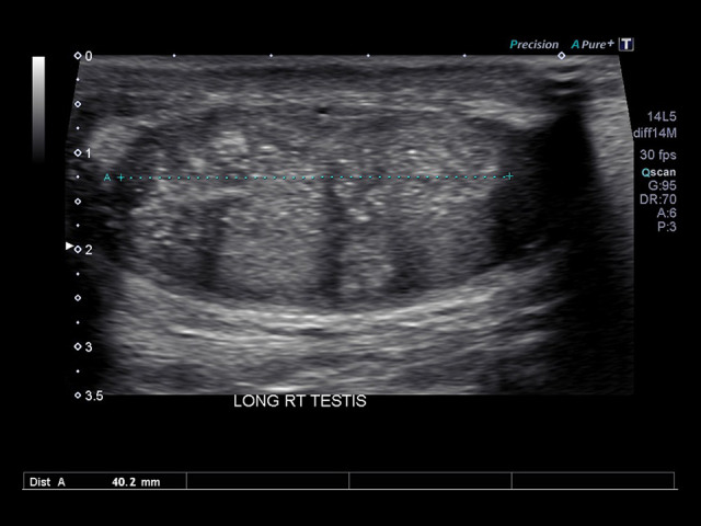
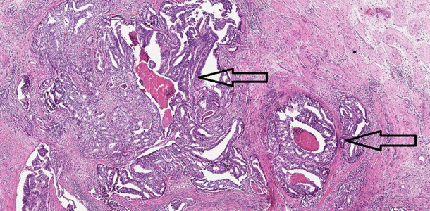
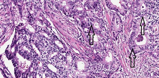
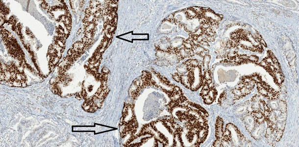
BACKGROUND Prostate cancer is the most common non-cutaneous cancer in men. While approximately three-quarters of all cases present as localized disease, the rate of metastatic disease has been increasing. Common sites of metastatic prostate cancer include regional lymph nodes, bones, and lungs. In this case report, we discuss a man with a history of low-risk prostate cancer who developed a testicular mass, which was ultimately diagnosed as a solitary testicular metastasis. CASE REPORT An abnormal nodule on the left apex area was identified on a digital rectal exam of an otherwise healthy 67-year-old man in February 2008. The patient underwent an ultrasound-guided transrectal biopsy of the prostate gland in April 2008. The biopsy demonstrated adenocarcinoma of the prostate, Gleason 6 (3+3), with tumor present in 3 out of 12 submitted cores in up to 20% of biopsy specimens. Following treatment, his prostate cancer remained quiescent for several years. He was also found to have a urethral bulbar stricture that required dilation; during the procedure, a nurse detected an abnormality in the right testicle while prepping the patient. A follow-up testicular ultrasound in September 2020 identified an abnormal heterogeneous area with calcifications within the right testicle. Following radical right orchiectomy, pathology revealed metastatic prostatic adenocarcinoma, acinar type, with lymphovascular invasion present at the spermatic cord margin. CONCLUSIONS Surveillance for prostate cancer following treatment, even for low-risk disease, should always be continued. Although rare, recurrence and metastasis can occur in patients with low and even absent post-treatment prostate-specific antigen levels.
Conflict of interest statement
Figures
References
-
- Dutt N, Bates AW, Baithun SI. Secondary neoplasms of the male genital tract with different patterns of involvement in adults and children. Histopathology. 2000;37(4):323–31. - PubMed
-
- Wang G. Metastatic carcinoma to the testis – a mini review. Journal of Rare Diseases Research & Treatment. 2019;4(2):16–22.
-
- Vitolo U, Ferreri AJM, Zucca E. Primary testicular lymphoma. Crit Rev Oncol Hematol. 2008;65(2):183–89. - PubMed
-
- Greene KL, Albertsen P, Babaian RJ, et al. Prostate specific antigen best practice statement: 2009 update. J Urol. 2009;182(5):2232–41. - PubMed
Publication types
MeSH terms
Substances
LinkOut - more resources
Full Text Sources
Medical
Miscellaneous
