. 2022 Mar 14;13(1):1305.
doi: 10.1038/s41467-022-28902-8.

An achromatic X-ray lens

Affiliations

An achromatic X-ray lens

Adam Kubec et al. Nat Commun. .

Abstract

Diffractive and refractive optical elements have become an integral part of most high-resolution X-ray microscopes. However, they suffer from inherent chromatic aberration. This has to date restricted their use to narrow-bandwidth radiation, essentially limiting such high-resolution X-ray microscopes to high-brightness synchrotron sources. Similar to visible light optics, one way to tackle chromatic aberration is by combining a focusing and a defocusing optic with different dispersive powers. Here, we present the first successful experimental realisation of an X-ray achromat, consisting of a focusing diffractive Fresnel zone plate (FZP) and a defocusing refractive lens (RL). Using scanning transmission X-ray microscopy (STXM) and ptychography, we demonstrate sub-micrometre achromatic focusing over a wide energy range without any focal adjustment. This type of X-ray achromat will overcome previous limitations set by the chromatic aberration of diffractive and refractive optics and paves the way for new applications in spectroscopy and microscopy at broadband X-ray tube sources.


Conflict of interest statement

A.K. is an employee of XRnanotech GmbH. The remaining authors declare no competing interests.

Figures

Fig. 1
Fig. 1. Concept of the X-ray achromat and experimental setup.
a Principle of achromatic focusing: The chromaticity of the defocusing refractive lens (RL) acts as a corrector for the chromatic behaviour of the focusing Fresnel zone plate (FZP). b Scanning electron microscopy (SEM) image of a nickel FZP fabricated by electron-beam lithography and nickel electroplating, as used for the comparison measurements. c SEM image of the RL consisting of four stacked paraboloids 3D-printed using two-photon polymerisation lithography. d Sketch of the experimental setup for scanning transmission X-ray microscopy (STXM) and ptychography using the achromat as a focusing optic.
Fig. 2
Fig. 2. Demonstration of STXM imaging at different energies using the achromat.
a STXM images of the Siemens star sample shown in panel b obtained with the achromat, indicating an achromatic range of  > 1 keV around the optimum energy of ~ 6.4 keV. b SEM image of the Siemens star test sample. The radial lines and spaces (L/S) at the outer and inner concentric rings have widths of 400 nm and 200 nm, respectively, see red arrows. c Comparison of the STXM results in the energy range of 6.0 keV to 6.4 keV obtained with the achromat (top) and the conventional FZP (bottom). While the contrast of the FZP images changes rapidly with the energy, the image quality achieved with the achromat varies only little.
Fig. 3
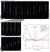
Fig. 3. Evolution of the X-ray beam profile with the energy measured with ptychography.
a Caustics at energies from 5.2 keV to 8.0 keV obtained with the achromat. The red dashed line indicates the location of the focal plane at the different energies. b Comparison of the caustics obtained with the achromat and the FZP. While the position of the focal plane remains almost constant with the energy for the achromat (red dashed line), it changes rapidly for the FZP (blue dashed line). c Calculated curves (solid and dashed lines) and experimental data (dots) for the focal length versus energy for the FZP (blue) and the achromat (red; solid: based on Eq. (6), dashed: based on tabulated refractive index values for the calculation of fRL at each energy).
Fig. 4
Fig. 4. Simulations of polychromatic X-ray focusing with an achromatic lens and with a single FZP (energy range from 5.6 keV to 6.8 keV).
a Convolution of the simulated polychromatic X-ray beam of the achromat with the binarised image of the Siemens star in Fig. 2b. b Convolution of the same image with the simulated polychromatic X-ray beam of the FZP. Gaussian noise was added to both images after convolution to model the image noise in experimental data. c Line profiles of the simulated polychromatic X-ray beams (normalised to central peak) for the achromat (red) and the FZP (blue).

References

    1. Schroer C, Lengeler B. X-Ray Optics. New York: Springer New York; 2007.
    1. Salditt, T. & Osterhoff, M. X-ray Focusing and Optics. (Springer International Publishing, Cham, 2020).
    1. Jacobsen, C. X-ray Microscopy, 199–240. Chapter 5: X-Ray Focusing Optics (Cambridge University Press, Cambridge, 2019).
    1. Wolter H. Spiegelsysteme streifenden Einfalls als abbildende Optiken für Röntgenstrahlen. Ann. Phys. 1952;445:94–114. doi: 10.1002/andp.19524450108. - DOI
    1. Matsuyama S, et al. 50-nm-resolution full-field X-ray microscope without chromatic aberration using total-reflection imaging mirrors. Sci. Rep. 2017;7:46358. doi: 10.1038/srep46358. - DOI - PMC - PubMed