. 2022 Apr;21(4):e13582.
doi: 10.1111/acel.13582. Epub 2022 Mar 15.

Quantitative restoration of immune defense in old animals determined by naive antigen-specific CD8 T-cell numbers

Affiliations

Quantitative restoration of immune defense in old animals determined by naive antigen-specific CD8 T-cell numbers

Jennifer L Uhrlaub et al. Aging Cell. 2022 Apr.

Abstract

Older humans and animals often exhibit reduced immune responses to infection and vaccination, and this often directly correlates to the numbers and frequency of naive T (Tn) cells. We found such a correlation between reduced numbers of blood CD8+ Tn cells and severe clinical outcomes of West Nile virus (WNV) in both humans naturally exposed to, and mice experimentally infected with, WNV. To examine possible causality, we sought to increase the number of CD8 Tn cells by treating C57BL/6 mice with IL-7 complexes (IL-7C, anti-IL-7 mAb bound to IL-7), shown previously to efficiently increase peripheral T-cell numbers by homeostatic proliferation. T cells underwent robust expansion following IL-7C administration to old mice increasing the number of total T cells (>fourfold) and NS4b:H-2Db -restricted antigen-specific CD8 T cells (twofold). This improved the numbers of NS4b-specific CD8 T cells detected at the peak of the response against WNV, but not survival of WNV challenge. IL-7C-treated old animals also showed no improvement in WNV-specific effector immunity (neutralizing antibody and in vivo T-cell cytotoxicity). To test quantitative limits to which CD8 Tn cell restoration could improve protective immunity, we transferred graded doses of Ag-specific precursors into old mice and showed that injection of 5400 (but not of 1800 or 600) adult naive WNV-specific CD8 T cells significantly increased survival after WNV. These results set quantitative limits to the level of Tn reconstitution necessary to improve immune defense in older organisms and are discussed in light of targets of immune reconstitution.

Keywords: IL-7/ CD8 T cells; immune aging; immune rejuvenation.


Conflict of interest statement

J.N.Ž. is co‐chair of the scientific advisory board of and receives research funding from Young Blood Institute, Inc.

Figures

FIGURE 1
FIGURE 1
Older adults experience more severe WNV disease and have a dearth of naive CD8 T cells. A cohort of 52 patients with WNV disease severity clinically classified as asymptomatic, WN fever (WNF), WN meningitis (WNM), and WN meningitis and encephalitis (WNME) were retrospectively recruited to participate in our study. The age of the patients experiencing the most severe disease is significantly older than asymptomatic (a). Naive (CD95low and CD28int/high, also confirmed additionally to be CCR7hi and CD45RA+) CD4 and CD8 T cells were phenotyped by FCM, and counts were extrapolated from clinical cell blood count (CBC) data collected on the fresh blood sample (b and c). Statistical significance evaluated by Kruskal–Wallis with Dunn's multiple comparisons test between all groups, +SEM. *≤ 0.05; **≤ 0.01; ***≤ 0.001
FIGURE 2
FIGURE 2
Decreased numbers of naive CD8 T cells in both young and old mice correlate with increased susceptibility to WNV. Adult (3–5 months) and old (19–22 months) C57BL/6 mice were infected with WNV, 1000pfu, f.p.; bled on Day 7 for FCM; and followed for susceptibility (a, schema). Absolute numbers of naive CD8 or CD4 T cells were enumerated per mL of blood on Day 7 post‐infection by FCM (b, c). Data compiled from two independent experiments. Statistical significance evaluated by t‐test, median. *≤ 0.05; **≤ 0.01; ****≤ 0.0001
FIGURE 3
FIGURE 3
IL‐7C treatment expands the total number of T cells available in secondary lymphoid tissues. Old (19–22 months) C57BL/6 mice were treated with IL‐7C on Days 1, 3, and 5 of the experiment and sacrificed on Days 7, 10, and 14 post‐first treatment alongside untreated adult (3–5 months) and old (19–22 months) (schema, a). The total number of lymphocytes (b), CD4 T cells (c), and CD8 T cells (d) are increased at indicated day(s) post‐first treatment. The total number of naive, CD44 low and CD62L high, CD4 and CD8 T cells are not significantly increased at any day post‐treatment (e, f). The expansion of CD4 T cells is not dominated by either CM (CD44 high, CD62L high, and CD49d high) or virtual memory (CD44 high, CD62L high, and CD49d low) phenotyped cells (g, h). Both CM and VM phenotype CD8 T cells are expanded at Day 10 post‐first treatment (i, j). WNV‐specific CD8 CTLp (Db‐NS4b2488) are not reliably increased by IL‐7C treatment (k). (b–j) Data shown are representative of two experiments with similar results and statistical significance evaluated by one‐way ANOVA with Dunn's multiple comparisons test. All comparisons are to untreated old mice. *≤ 0.05; **≤ 0.01. (k) IL‐7C‐treated group is Day 13 post‐first treatment. Data compiled from three independent experiments and statistical significance evaluated by t‐test comparison to O, median. *≤ 0.05
FIGURE 4
FIGURE 4
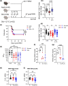
IL‐7C treatment of old mice improves WNV‐specific CD8 T‐cell responses without impacting survival. Mice of the following ages and treatments: Adult (3–5 months), Old (19–22 months), and Old treated with IL‐7C 13 days before challenge (19–22 months); were infected with WNV (1000pfu, f.p.; n = 45–52 per group). On Day 7 post‐WNV infection, mice are bled and monitored for survival (schema, a). Number of WNV‐specific CD8 T cells per mL of blood on Day 7 post‐WNV (b). WNV survival at Day 45 post‐infection (c). Number of WNV‐specific CD8 T cells from (b) broken by those that survived and those that died from WNV in each group (c). Neutralizing antibody titers on Day 7 post‐WNV broken by those that survived and those that from died from WNV in each group (e). In vivo killing assay on Day 7 post‐WNV for NS4b2488 (f) and ENV641 (g). Total WNV‐ENV and DIII‐specific memory antibody levels >45 days post‐infection (Figure h, i). Data compiled from two experiments. Statistical significance evaluated by ANOVA in (b, f, g) and t‐test in (d) and (e) as represented on graphs. Significance of survival evaluated by Log‐rank (Mantel‐Cox) test. *≤ 0.05; **≤ 0.01; ***≤ 0.001; ****≤ 0.0001.
FIGURE 5
FIGURE 5
IL7‐C treatment does not induce T‐cell exhaustion. Adult (3–5 months) and old (19–22 months) mice were treated with IL‐7C 13 days before infection with WNV (1000pfu, f.p.). On Day 7 post‐WNV infection, mice were bled and circulating PBMCs were analyzed by FCM for expression of PD1, CD244 (2B4), and CD223 (LAG‐3) on NS4b2488+ CD8 T cells (a–c). Data compiled from two experiments. Statistical significance evaluated by ANOVA in (all groups) and t‐test in (by age) as represented on graphs. *≤ 0.05; **≤ 0.01; ***≤ 0.001
FIGURE 6
FIGURE 6
Adoptive transfer of WNV‐I Tg CD8 T cells rescues old mice from WNV susceptibility. WNV‐I CD8 T cells (5400, 1600, or 600) were transferred to old mice (19‐22 months) the day before WNV challenge (1000pfu, f.p.). On Day 7 post‐infection, mice were bled and total numbers of circulating WNV‐specific CD8 T cells were enumerated per mL of blood (a). Mice were monitored for survival to Day 45 post‐infection (b). The frequency of donor WNV‐I CD8 T cells (CD45.1) contributing to the response in (a) is shown in (c). In vivo killing assay on Day 7 post‐WNV for NS4b2488 (d). Data compiled (a–c) or representative (d) of two experiments. Statistical significance evaluated by ANOVA (a, c, d) with multiple comparisons test. Significance of survival evaluated by Log‐rank (Mantel‐Cox) test. *≤ 0.05; **≤ 0.01; ***≤ 0.001; ****≤ 0.0001

References

    1. Aguilar‐Valenzuela, R. , Netland, J. , Seo, Y. J. , Bevan, M. J. , Grakoui, A. , & Suthar, M. S. (2018). Dynamics of tissue‐specific CD8(+) T cell responses during west nile virus infection. Journal of Virology, 92(10), 14–18. 10.1128/jvi.00014-18 - DOI - PMC - PubMed
    1. Blattman, J. N. , Antia, R. , Sourdive, D. J. D. , Wang, X. , Kaech, S. M. , Murali‐Krishna, K. , Altman, J. D. , & Ahmed, R. (2002). Estimating the precursor frequency of naive antigen‐specific CD8 T cells. Journal of Experimental Medicine, 195(5), 657–664. 10.1084/jem.20001021 - DOI - PMC - PubMed
    1. Boyman, O. , Kovar, M. , Rubinstein, M. P. , Surh, C. D. , & Sprent, J. (2006). Selective stimulation of T cell subsets with antibody‐cytokine immune complexes. Science, 311(5769), 1924–1927. 10.1126/science.1122927 - DOI - PubMed
    1. Boyman, O. , Ramsey, C. , Kim, D. M. , Sprent, J. , & Surh, C. D. (2008). IL‐7/anti‐IL‐7 mAb complexes restore T cell development and induce homeostatic T Cell expansion without lymphopenia. The Journal of Immunology, 180(11), 7265–7275. 10.4049/jimmunol.180.11.7265 - DOI - PubMed
    1. Brien, J. D. , Uhrlaub, J. L. , Hirsch, A. , Wiley, C. A. , & Nikolich‐Zugich, J. (2009). Key role of T cell defects in age‐related vulnerability to West Nile virus. Journal of Experimental Medicine, 206(12), 2735–2745. 10.1084/jem.20090222 - DOI - PMC - PubMed

Publication types