

Image Quality and Lesion Detectability of Lower-Dose Abdominopelvic CT Obtained Using Deep Learning Image Reconstruction
- PMID: 35289146
- PMCID: PMC8961013
- DOI: 10.3348/kjr.2021.0683
Image Quality and Lesion Detectability of Lower-Dose Abdominopelvic CT Obtained Using Deep Learning Image Reconstruction
Abstract
Objective: To evaluate the image quality and lesion detectability of lower-dose CT (LDCT) of the abdomen and pelvis obtained using a deep learning image reconstruction (DLIR) algorithm compared with those of standard-dose CT (SDCT) images.
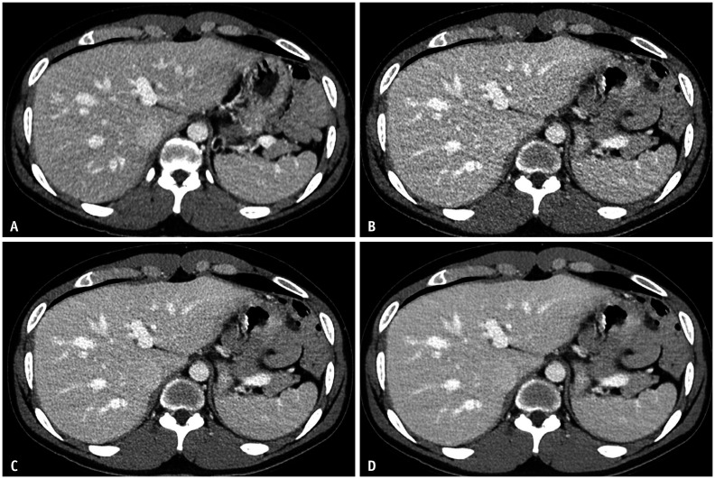
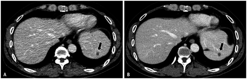
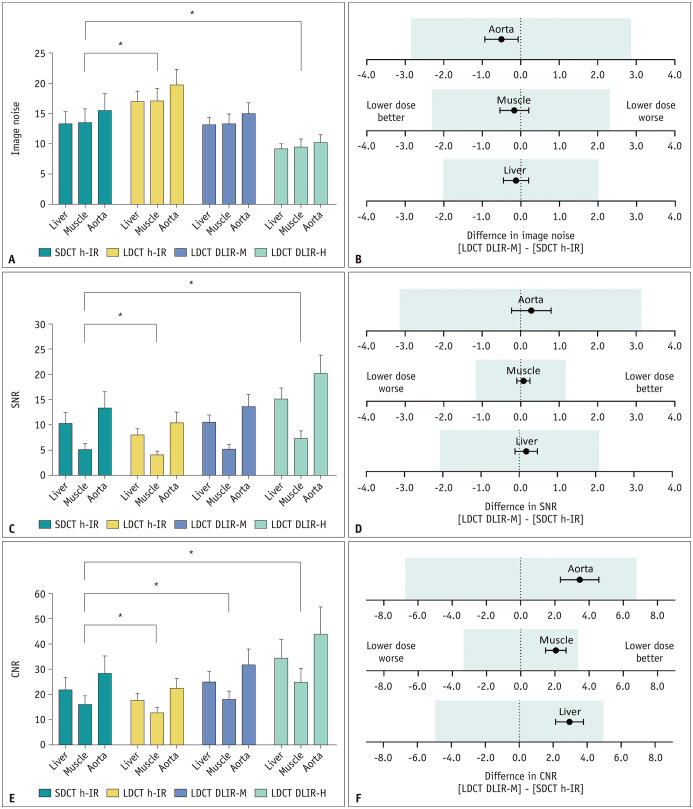
Materials and methods: This retrospective study included 123 patients (mean age ± standard deviation, 63 ± 11 years; male:female, 70:53) who underwent contrast-enhanced abdominopelvic LDCT between May and August 2020 and had prior SDCT obtained using the same CT scanner within a year. LDCT images were reconstructed with hybrid iterative reconstruction (h-IR) and DLIR at medium and high strengths (DLIR-M and DLIR-H), while SDCT images were reconstructed with h-IR. For quantitative image quality analysis, image noise, signal-to-noise ratio, and contrast-to-noise ratio were measured in the liver, muscle, and aorta. Among the three different LDCT reconstruction algorithms, the one showing the smallest difference in quantitative parameters from those of SDCT images was selected for qualitative image quality analysis and lesion detectability evaluation. For qualitative analysis, overall image quality, image noise, image sharpness, image texture, and lesion conspicuity were graded using a 5-point scale by two radiologists. Observer performance in focal liver lesion detection was evaluated by comparing the jackknife free-response receiver operating characteristic figures-of-merit (FOM).
Results: LDCT (35.1% dose reduction compared with SDCT) images obtained using DLIR-M showed similar quantitative measures to those of SDCT with h-IR images. All qualitative parameters of LDCT with DLIR-M images but image texture were similar to or significantly better than those of SDCT with h-IR images. The lesion detectability on LDCT with DLIR-M images was not significantly different from that of SDCT with h-IR images (reader-averaged FOM, 0.887 vs. 0.874, respectively; p = 0.581).
Conclusion: Overall image quality and detectability of focal liver lesions is preserved in contrast-enhanced abdominopelvic LDCT obtained with DLIR-M relative to those in SDCT with h-IR.
Keywords: Abdomen; Computed tomography; Deep learning; Image reconstruction; Radiation dose.
Copyright © 2022 The Korean Society of Radiology.
Conflict of interest statement
Yong Eun Chung who is on the editorial board of the
Figures

References
-
- Huang L, Snyder AR, Morgan WF. Radiation-induced genomic instability and its implications for radiation carcinogenesis. Oncogene. 2003;22:5848–5854. - PubMed
-
- Sodickson A, Baeyens PF, Andriole KP, Prevedello LM, Nawfel RD, Hanson R, et al. Recurrent CT, cumulative radiation exposure, and associated radiation-induced cancer risks from CT of adults. Radiology. 2009;251:175–184. - PubMed
-
- Mullenders L, Atkinson M, Paretzke H, Sabatier L, Bouffler S. Assessing cancer risks of low-dose radiation. Nat Rev Cancer. 2009;9:596–604. - PubMed
-
- National Research Council. Health risks from exposure to low levels of ionizing radiation: BEIR VII phase 2. Washington DC: National Academies Press; 2006. p. 406. - PubMed
MeSH terms
LinkOut - more resources
Full Text Sources
