. 2022 Mar 15;132(6):e128808.
doi: 10.1172/JCI128808.

Genetically programmed alternative splicing of NEMO mediates an autoinflammatory disease phenotype

Affiliations

Genetically programmed alternative splicing of NEMO mediates an autoinflammatory disease phenotype

Younglang Lee et al. J Clin Invest. .

Abstract

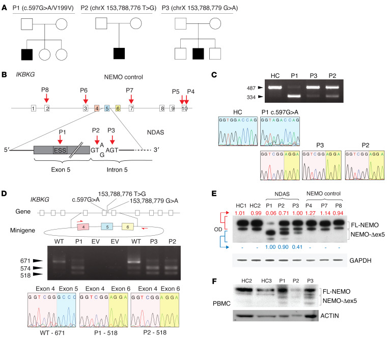
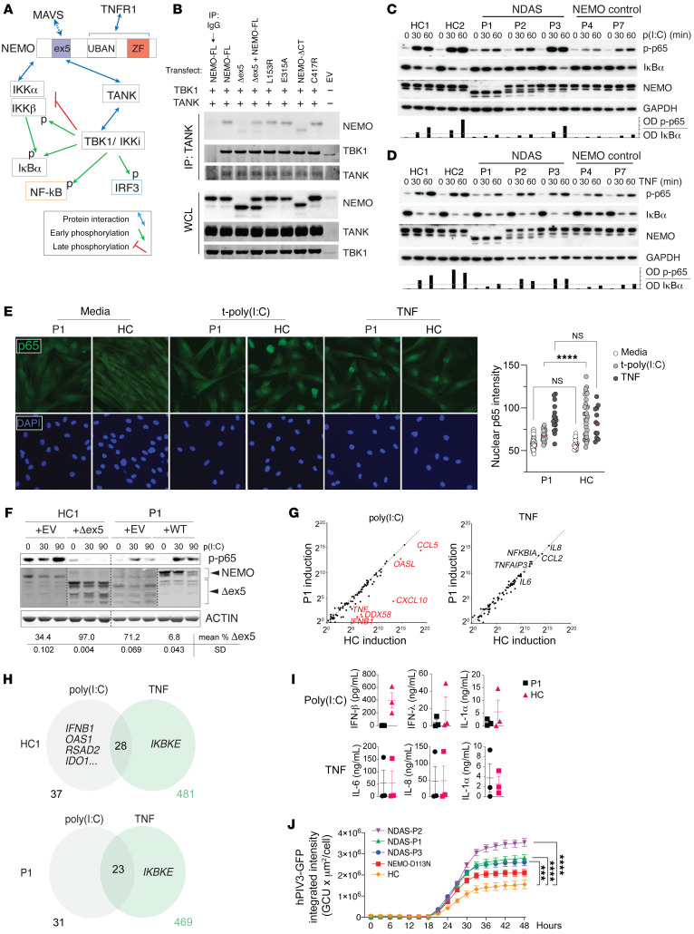
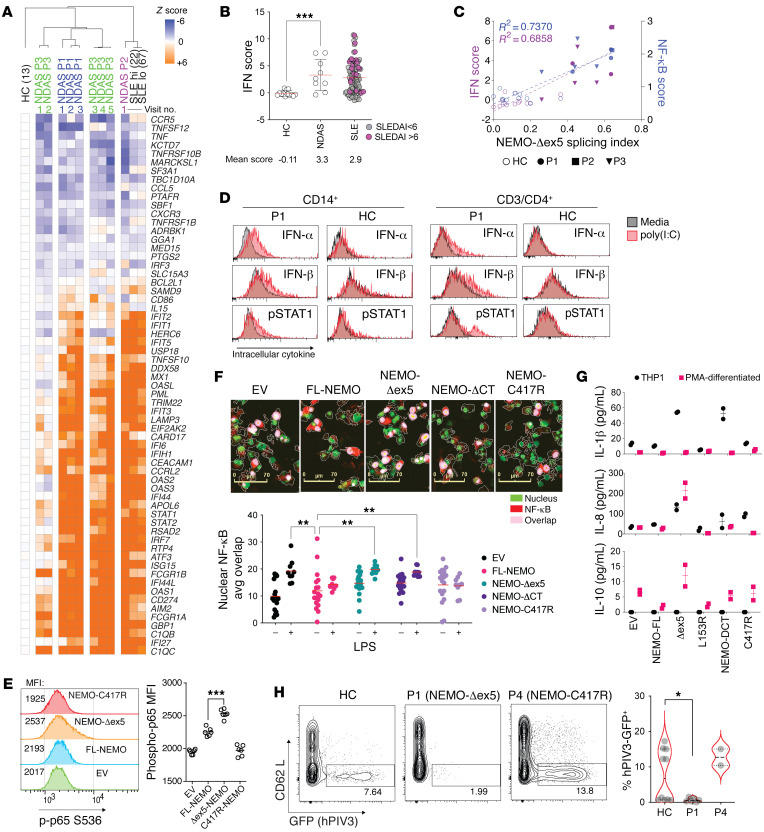
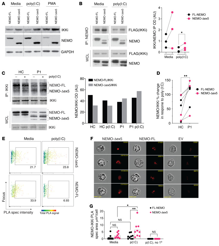
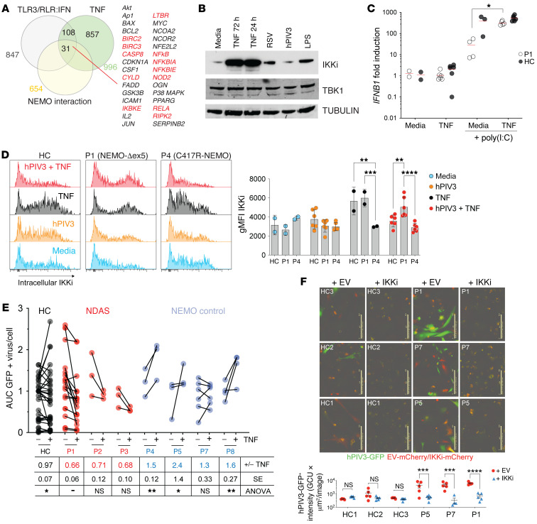
Host defense and inflammation are regulated by the NF-κB essential modulator (NEMO), a scaffolding protein with a broad immune cell and tissue expression profile. Hypomorphic mutations in inhibitor of NF-κB kinase regulatory subunit gamma (IKBKG) encoding NEMO typically present with immunodeficiency. Here, we characterized a pediatric autoinflammatory syndrome in 3 unrelated male patients with distinct X-linked IKBKG germline mutations that led to overexpression of a NEMO protein isoform lacking the domain encoded by exon 5 (NEMO-Δex5). This isoform failed to associate with TANK binding kinase 1 (TBK1), and dermal fibroblasts from affected patients activated NF-κB in response to TNF but not TLR3 or RIG-I-like receptor (RLR) stimulation when isoform levels were high. By contrast, T cells, monocytes, and macrophages that expressed NEMO-Δex5 exhibited increased NF-κB activation and IFN production, and blood cells from these patients expressed a strong IFN and NF-κB transcriptional signature. Immune cells and TNF-stimulated dermal fibroblasts upregulated the inducible IKK protein (IKKi) that was stabilized by NEMO-Δex5, promoting type I IFN induction and antiviral responses. These data revealed how IKBKG mutations that lead to alternative splicing of skipping exon 5 cause a clinical phenotype we have named NEMO deleted exon 5 autoinflammatory syndrome (NDAS), distinct from the immune deficiency syndrome resulting from loss-of-function IKBKG mutations.

Trial registration: ClinicalTrials.gov NCT00001788 NCT00001372 NCT02974595.

Keywords: Genetic diseases; Genetics; Immunology; Inflammation; Innate immunity; Signal transduction.


Conflict of interest statement

Conflict of interest: CE has served on an advisory board for Union Chimique Belge. RGM has received study support under government cooperative research and development agreements (CRADAs) from Swedish Orphan Biovitrum AB, Regeneron, Eli Lilly, and Novartis. RMS has been an employee of Novartis since 2018 and currently owns Novartis stock, though he worked for the NIH at the time he worked on this manuscript. EPH’s spouse receives income from Eli Lilly and holds unvested restricted stock options.

Figures

Figure 1
Figure 1. A synonymous exonic mutation and 2 intronic splice donor mutations mediate alternative splicing of IKBKG and lead to severe autoinflammatory disease.
(A) Pedigree and clinical features of developmental and inflammatory diseases in patients P1–P3. (B) Schema of mutations identified in patients P1–P3 and other NEMO mutants. Note exon coloring used for C and D. Patient P4 has the well-described NEMO-C417R zinc finger hypomorphic mutation. I DNA gel electrophoresis of IKBKG cDNA from P1, P2, P3, and healthy control (HC) dermal fibroblasts. Chromatograms of Sanger-sequenced FL isoforms from P1 and HCs and the variant isoform from P1–P3. (D) Schema of minigene spanning IKBKG exons 4–6 containing the reference sequence or patient mutations. DNA electrophoresis of cDNA and sequence traces are shown as In C. The 574 bp fragment contains noncoding intronic sequence in addition to a fragment of exon 5. WT = IKBKG minigene containing reference sequence. (E) Western blot of NEMO from patients P1–P3 and NEMO control patient skin fibroblasts, optical densitometry of FL-NEMO relative to loading control is shown in red, of NEMO-Δex5 in blue. (F) Western blot of PBMCs and whole cell lysate from patients P1, P2, and P3 and 2 healthy controls (HCs).
Figure 2
Figure 2. NEMO-Δex5 blocks NF-κB activation in response to poly(I:C) but TNF-induced NF-κB remains intact.
(A) NEMO/TANK/TBK1 interaction and function. (B) HEK293T cells transfected with TANK, TBK1, and NEMO followed by TANK IP and Western blot as indicated. (C and D) Patient and healthy control (HC) dermal fibroblasts (DFs) were stimulated with poly(I:C) in a time course followed by Western blot as indicated. Quantitation of phospho-p65/IκBα by optical densitometry, below. Dashed line at 20%. (E) P1 and HC DFs were stimulated with TNF or poly(I:C) for 60 minutes and stained for NF-κB p65 visualized at 200× magnification by immunofluorescence (Keyence BZ9000), quantitation of p65 nuclear intensity, right. Statistical significance by 2-way ANOVA with Tukey’s multiple-comparison test. (F) DFs from HC and P1 were stably transduced with NEMO-Δex5 or FL-NEMO, respectively, and stimulated with poly(I:C) in a time course; lysates probed by Western blot as indicated. Different exposure intensity of NEMO panels (HC/EV, HC/Δex5, P1/EV, and P1/WT) was required to reveal all relevant bands, relative quantitation by optical densitometry, below. (G) P1 and HC DFs stimulated with poly(I:C), TNF, or cultured in media for 3 hours. Average gene expression by NanoString, ratio in stimulated/untreated from n = 3 independent experiments. (H) DFs treated as in G with relevant differentially expressed genes by RNA-Seq and totals shown, n = 3 technical replicates per media and TNF condition, n = 2 for poly(I:C) treatment. (I) P1 and HC DFs stimulated with TNF or poly(I:C) for 12 hours, cytokine production by capture bead assay, representative experiment of n = 3. (J) DFs from patients with NDAS, NEMO controls, and HCs incubated with hPIV3-GFP. Quantitation of total GFP intensity per cell, error bar indicates SEM. Two-way ANOVA applying Dunnett’s multiple-comparison test for adjusted P values, pertaining to 48-hour point. ***P < 0.001; ****P < 0.0001. Poly(I:C) dose 10 μg/mL and TNF 20 ng/mL throughout. All results representative of at least 2 independent experiments.
Figure 3
Figure 3. Increased NF-κB activation and type I IFN production in T cells and monocytes expressing NEMO-Δex5.
(A) Relative gene expression by whole-blood RNA-Seq from P1, P2, and P3 obtained at serial clinic visits; patients with systemic lupus erythematosus (SLE) with SLEDAI greater than 6 (SLE hi, n = 22); SLEDAI less than 6 (SLE lo, n = 76), and from healthy controls (n = 13). (B) IFN signature gene score (see Methods) from whole-blood RNA-Seq data, ordinary 1-way ANOVA, Dunnett’s correction for multiple comparisons, group mean indicated by red bar with SD. (C) IFN and NF-κB scores plotted versus NEMO-Δex5 splicing index, least squares linear regression fit, R2 shown. (D) P1 and HC PBMCs cultured in media or stimulated with poly(I:C) 10 μg/mL for 60 minutes and treated with brefeldin for 4 hours, stained with antibodies against IFN-α, IFN-β, p-STAT1, CD14, and CD4. (E) THP1 were stably transduced to express FL or mutant forms of NEMO. NF-κB p65 phosphorylation was measured by intracellular flow cytometry. (F) PMA-differentiated reconstituted THP1 cells were stimulated with LPS 10 ng/mL for 60 minutes, fixed, and stained with anti–NF-κB p65 (red) and nuclear dye (green). Scale bars: 70 μm. Areas of nuclear p65 overlap (pink) were quantitated (below). Means compared by 2-way ANOVA with Dunnett’s correction. (G) Secreted cytokines in cell culture supernatant from NEMO-reconstituted THP1 cells (black symbols) after 72-hour PMA differentiation (red). (H) P1, P4, and HC blasting T cells were infected with GFP-expressing human parainfluenza virus 3 (hPIV3-GFP) for 48 hours; n = 6, 2, and 10 replicates, respectively. Frequency of hPIV3-GFP+ cells, right. P value calculated with Mann-Whitney unpaired, 2-tailed U test. All data except from Figures 3A–C are representative of at least 2 independent experiments. *P < 0.05; **P < 0.01; ***P < 0.001.
Figure 4
Figure 4. NEMO-Δex5 and IKKi form a stable complex in response to poly(I:C) stimulation.
(A) THP1 cells reconstituted with FL and mutant NEMO forms and stimulated with poly(I:C) for 90 minutes or differentiated with PMA for 72 hours followed by Western blot of whole cell lysates. (B) HEK293T cells were transfected with FL or NEMO-Δex5 followed by stimulation with poly(I:C) 10 μg/mL for 8 hours and IP of IKKi using FLAG epitope. The membrane was probed for IKKi and NEMO with quantitation of pulled down IKKi normalized to NEMO on the right; means compared by paired, 2-tailed t test. (C) HC and P1 blasting T cells stimulated with poly(I:C) 10 μg/mL for 3 hours and IKKi isolated by IP. Western blot to detect co-immunoprecipitated NEMO, and IKKi blot as IP control, (right) quantitation of NEMO pulldown with IKKi by densitometry, black bars correspond to FL-NEMO bands and gray bars to NEMO-Δex5 forms in top panel (ImageJ). (D) Quantitation of total NEMO pulldown (NEMO-FL + NEMO-Δex5) as in C, shown as poly(I:C) response [ratio of poly(I:C) treated/media] (n = 4 independent IPs from NDAS-P1 and 4 IPs from HC T cells; means compared by paired, 2-tailed t test. Lines link paired data obtained from the same experiment. (E) NEMO-reconstituted THP1 cells in media or stimulated with LMW poly(I:C) 10 μg/mL for 60 minutes with NEMO and IKKi proximity ligation assay (PLA) spec intensity (x axis) and total NEMO/IKKi complex intensity (rainbow heatmap) using specific antibodies against IKKi and NEMO. (F) Four representative images taken at 40× original magnification of cells treated for 60 minutes as in E, with average signal area quantified; means compared by 2-way ANOVA with Tukey’s correction (G). EV, empty vector; FL, full length; no 1°, omission of primary antibody control. All results representative of at least 2 independent experiments. *P < 0.05; **P < 0.01.
Figure 5
Figure 5. IKKi can be induced by TNF in hPIV3-infected P1 dermal fibroblasts, Ih rescues innate antiviral immunity.
(A) Venn diagram indicates the intersection of TNF-inducible, TLR3/RLR/IRF3 pathway genes that interact with NEMO, both directly (red) and indirectly (black). (B) IKKi expression in primary dermal fibroblasts from HCs in response to TNF 20 ng/mL for 24 hours or TNF 20 ng/mL, LPS 10 ng/mL, or viral infection for 72 hours detected by Western blot. TBK1 and TUBULIN served as loading controls. (C) P1 and HC skin fibroblasts were stimulated with poly(I:C) 10 μg/mL for 3 hours with or without TNF 20 ng/mL costimulation, and IFNB1 gene expression was determined by qPCR, 2-tailed Mann-Whitney U test. (D) P1, P4, and HC dermal fibroblasts were infected with hPIV3-GFP and treated with TNF 20 ng/mL as indicated by red shaded histograms. At 72 hours, cells were fixed and stained intracellularly with antibody to detect IKKi expression (quantitated on far right). (E) NDAS, NEMO control, and HC dermal fibroblasts (or iPSC-derived fibroblast-like cells from P5) were infected with hPIV3-GFP with or without TNF 20 ng/mL costimulation and imaged in time course experiments of 48 to 72 hours duration to measure virus protein expression. AUC of total GFP intensity per cell was measured and within-experiment normalization to HC samples was performed. For HC and NDAS-P1 samples, n = 7 independent experiments, for all other samples n = 3 independent experiments, with total numbers of conditions shown; “+/- TNF” in figure table underneath indicates mean hPIV3-GFP AUC ratio of TNF-stimulated versus unstimulated infected cells. Means compared by 2-way ANOVA with Tukey’s multiple-comparison test. (F) iPSC-derived fibroblast-like cells were transduced with lentiviral overexpression vector encoding IKKi-IRES-mCherry and infected with hPIV3-GFP. Scale bars: 400 μm. Two-way ANOVA with Bonferroni’s correction for multiple comparisons. Data are representative of at least 2 independent experiments. *P < 0.05; **P < 0.01; ***P < 0.001; ****P < 0.0001.

Comment in

References

    1. Zhao T, et al. The NEMO adaptor bridges the nuclear factor-kappaB and interferon regulatory factor signaling pathways. Nat Immunol. 2007;8(6):592–600. doi: 10.1038/ni1465. - DOI - PubMed
    1. Pomerantz JL, Baltimore D. NF-kappaB activation by a signaling complex containing TRAF2, TANK and TBK1, a novel IKK-related kinase. EMBO J. 1999;18(23):6694–6704. doi: 10.1093/emboj/18.23.6694. - DOI - PMC - PubMed
    1. Clark K, et al. Novel cross-talk within the IKK family controls innate immunity. Biochem J. 2011;434(1):93–104. doi: 10.1042/BJ20101701. - DOI - PubMed
    1. Bruni D, et al. A novel IRAK1-IKKε signaling axis limits the activation of TAK1-IKKβ downstream of TLR3. J Immunol. 2013;190(6):2844–2856. doi: 10.4049/jimmunol.1202042. - DOI - PubMed
    1. Audry M, et al. NEMO is a key component of NF-κB- and IRF-3-dependent TLR3-mediated immunity to herpes simplex virus. J Allergy Clin Immunol. 2011;128(3):610–617. doi: 10.1016/j.jaci.2011.04.059. - DOI - PMC - PubMed

Publication types

Associated data