. 2022 Apr 26;13(2):e0325121.
doi: 10.1128/mbio.03251-21. Epub 2022 Mar 15.

Copper Induces Protein Aggregation, a Toxic Process Compensated by Molecular Chaperones

Affiliations

Copper Induces Protein Aggregation, a Toxic Process Compensated by Molecular Chaperones

Lisa Zuily et al. mBio. .

Abstract

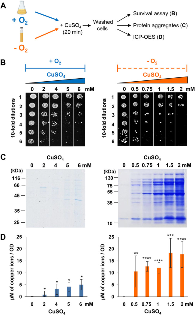
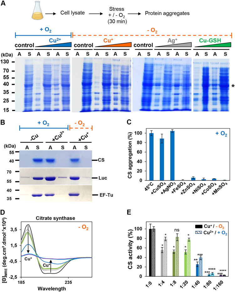
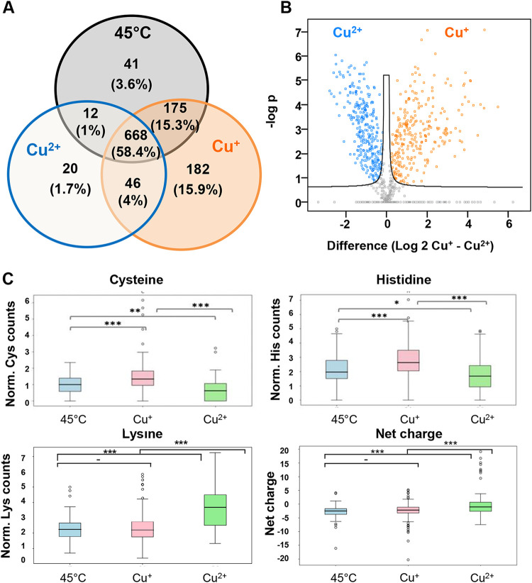
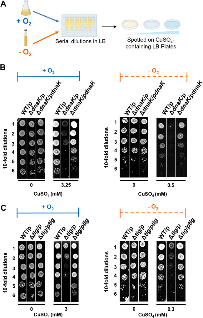
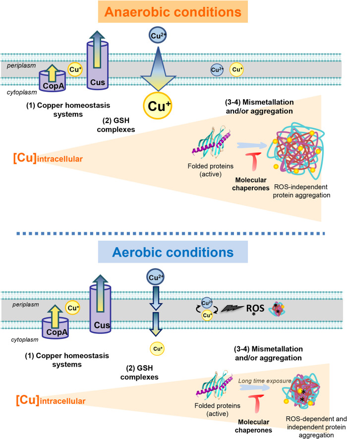
Copper is well known for its antimicrobial and antiviral properties. Under aerobic conditions, copper toxicity relies in part on the production of reactive oxygen species (ROS), especially in the periplasmic compartment. However, copper is significantly more toxic under anaerobic conditions, in which ROS cannot be produced. This toxicity has been proposed to arise from the inactivation of proteins through mismetallations. Here, using the bacterium Escherichia coli, we discovered that copper treatment under anaerobic conditions leads to a significant increase in protein aggregation. In vitro experiments using E. coli lysates and tightly controlled redox conditions confirmed that treatment with Cu+ under anaerobic conditions leads to severe ROS-independent protein aggregation. Proteomic analysis of aggregated proteins revealed an enrichment of cysteine- and histidine-containing proteins in the Cu+-treated samples, suggesting that nonspecific interactions of Cu+ with these residues are likely responsible for the observed protein aggregation. In addition, E. coli strains lacking the cytosolic chaperone DnaK or trigger factor are highly sensitive to copper stress. These results reveal that bacteria rely on these chaperone systems to protect themselves against Cu-mediated protein aggregation and further support our finding that Cu toxicity is related to Cu-induced protein aggregation. Overall, our work provides new insights into the mechanism of Cu toxicity and the defense mechanisms that bacteria employ to survive. IMPORTANCE With the increase of antibiotic drug resistance, alternative antibacterial treatment strategies are needed. Copper is a well-known antimicrobial and antiviral agent; however, the underlying molecular mechanisms by which copper causes cell death are not yet fully understood. Herein, we report the finding that Cu+, the physiologically relevant copper species in bacteria, causes widespread protein aggregation. We demonstrate that the molecular chaperones DnaK and trigger factor protect bacteria against Cu-induced cell death, highlighting, for the first time, the central role of these chaperones under Cu+ stress. Our studies reveal Cu-induced protein aggregation to be a central mechanism of Cu toxicity, a finding that will serve to guide future mechanistic studies and drug development.

Keywords: DnaK; Escherichia coli; copper homeostasis; copper stress; copper tolerance; heat shock; molecular chaperone; protein aggregation; proteostasis; stress response; trigger factor.


Conflict of interest statement

The authors declare no conflict of interest.

Figures

FIG 1
FIG 1
CuSO4 induces protein unfolding under anaerobic conditions in vivo. (A) E. coli cells were grown until they reached an OD600 of 0.7 and exposed to different concentrations of CuSO4 for 20 min at 37°C under aerobic (in blue) or anaerobic (in orange) conditions. Cells were washed several times to remove copper excess. They were subsequently analyzed by three different assays. (B) Three microliters of 10-fold serial dilutions was spotted onto LB agar plates in an aerobic (left panel) or anaerobic (right panel) atmosphere and incubated at 37°C. The plates are representative of at least three independent experiments. (C) Cells were lysed and after different centrifugation steps, aggregated proteins were isolated and loaded on 12% SDS-PAGE; the gels are representative of at least three independent experiments. (D) The amount of intracellular Cu content was measured by ICP-OES. Error bars represent standard deviations (SD) from triplicate experiments. Statistical analyses were performed using an unpaired two-tailed t test (*, P < 0,05; **, P < 0,01; ***, P < 0.001; ****, P < 0.0001), using their respective results from 0 mM treated cells as the comparative value.
FIG 2
FIG 2
In vitro inactivation, unfolding, and aggregation of proteins by Cu+ or Cu2+. (A) A 1-mg/mL concentration of E. coli lysate was incubated with or without 100 and 500 μM Cu2+ under aerobic conditions and Cu+, Ag+, or Cu-GSH under anaerobic conditions. Control samples correspond to the absence of copper; in the case of Cu+ treatment, the same amount of acetonitrile was added in the control samples. Aggregates (A) and soluble proteins (S) were separated by centrifugation and analyzed by 12% SDS-PAGE. An asterisk indicates EF-Tu protein identified by mass spectrometry. This experiment was performed in triplicate, and a representative result is shown. (B) A 2 μM concentration of citrate synthase (CS), luciferase (Luc), or EF-Tu was incubated for 30 min at 30°C in the absence of metal ions or in the presence of 80-fold molar excess of Cu2+ (under aerobic conditions) or Cu+ (under anaerobic conditions). Aggregates (A) and soluble proteins (S) were separated by centrifugation and analyzed by 12% SDS-PAGE. This experiment was performed in triplicate, and representative results are shown. (C) Aggregation of CS (2 μM) was measured by light scattering (360 nm) at 30°C, and end points were taken after 15 min of incubation with 80-fold molar excess of different metals under aerobic conditions. A value obtained at 30°C in the absence of metal ions was defined as 0%, and a value obtained after 15 min at 45°C corresponds to 100% of CS aggregation. Error bars represent standard deviation (SD). (D) Far-UV CD spectra of 2.5 μM CS treated without metal (black) or with a 4-fold (gray), 8-fold (dark green), 20-fold (light green), 40-fold (dark blue), 80-fold (light blue), or 160-fold (purple) molar excess of Cu+ (under anaerobic conditions) were recorded at 25°C. A representative result from several experiments is shown. (E) A 1.5 nM concentration of CS was incubated without metal (black) or with a 4-fold (gray), 8-fold (dark green), 20-fold (light green), 40-fold (dark blue), 80-fold (light blue), or 160-fold (purple) molar excess of Cu+ (under anaerobic conditions) or Cu2+ (same color code, with hatching, under aerobic conditions), at 25°C for 30 min, and CS-specific activity was monitored. CS-specific activity obtained without Cu addition represents 100% of CS activity. Error bars represent standard deviation (SD). Statistical analyses were performed with an unpaired two-tailed t test (*, P < 0.05; **, P < 0.01), using CS without metal as a comparative value.
FIG 3
FIG 3
Aggregation-prone proteins and changes in abundance of proteins/residues after different treatments. (A) Venn diagram of identified insoluble proteins by mass spectrometry analysis. Insoluble proteins obtained at 30°C were subtracted from all three sets. The number of proteins identified under each condition is depicted, with the total number of aggregates under all three conditions set to 100%. (B) Volcano plots represent comparative analysis of differential abundance of insoluble proteins obtained after two different treatments. Significantly abundant proteins are colored in either orange (Cu+) or blue (Cu2+), according to the FDR of 0.05 and a fold change greater than 2. The volcano plot is related to Table S1. (C) Box plots of amino acid propensities (cysteine, histidine, and lysine) or net charge of proteins aggregated following a treatment with either 45°C (blue), Cu+ (pink), or Cu2+ (green). The features were normalized to the protein length. PASTA 2.0 (26) was used for prediction of protein aggregation and disorder propensity. Instability and amino acid propensities were evaluated by using in-house scripts with Python methods from the website https://biopython.org/DIST/docs/api/Bio.SeqUtils.ProtParam.ProteinAnalysis-class.html#flexibility. Two-tailed Student's t tests with similar variance were used to evaluate statistically different features. Significantly different sets are marked according to P values of 0.001 (***), 0.01 (**), or 0.05 (*).
FIG 4
FIG 4
Gene response and role of molecular chaperones after short-term exposure to copper. (A) E. coli culture was exposed to different CuSO4 concentrations either anaerobically (orange) or aerobically (blue) for 20 min at 37°C (B) or 30°C (C). The cells were directly centrifuged to extract RNA from the WT strain (B) or serially diluted in LB and spotted on plates (C). In contrast to Fig. 1, no washing step was added because the cells were either quickly centrifuged (B) or diluted in LB, which quenches the excess of copper (C). (B) Quantitative RT-PCR analysis of different sets of genes encoding molecular chaperones (dnaK and tig) and involved in the heat shock response (dnaK), copper homeostasis (copA), or the oxidative stress response (sodA). The standard deviation is represented by error bars calculated from at least three independent experiments. (C) The WT or the ΔdnaK and Δtig mutant strains were stressed and grown under aerobic (blue) or anaerobic (orange) conditions. Plates were incubated at 30°C overnight, and the results are representative of at least three experiments.
FIG 5
FIG 5
Molecular chaperones protect cells against long-term exposure to copper under both aerobic and anaerobic conditions. (A) Strains were grown at 30°C until they reached an OD of 0.7 under aerobic (in blue) or anaerobic (in orange) conditions. Serial dilutions of these strains were spotted on LB agar supplemented or not with different concentrations of CuSO4 and also containing ampicillin and IPTG. Plates were incubated in an aerobic or anaerobic atmosphere overnight at 30°C. (B) The MC4100 WT strain (WT) and the ΔdnaK::CmrdnaK) mutant containing the empty vector pSE380 (p) or the plasmid expressing dnaK (pdnaK) were grown at 30°C with ampicillin. After following the procedure described for panel A, representative plates are depicted. (C) The MC4100 WT and Δtig::Cmr mutant strain containing the empty vector pSE380 (p) or the plasmid expressing tig (ptig) were grown at 30°C and then spotted on LB plates containing copper as described for panel A. Plates are representative of at least three experiments.
FIG 6
FIG 6
Model of CuSO4 effects on protein folding and chaperone function under aerobic or anaerobic growth conditions. Under non-stress conditions, proteins are well folded and active. Exposure of proteins to CuSO4 (Cu2+) under anaerobic conditions leads to the intracellular accumulation of Cu+ (arrow). Depending on the CuSO4 concentration and the incubation time, a gradual increase of intracellular copper content will occur (represented by the triangular orange shading). This will first induce the expression of copper homeostasis systems (1st phase). In this figure, only two systems are shown: the CopA and Cus systems. They are both known to export intracellular copper excess. Intracellular cytoplasmic copper will react with GSH (2nd phase), which will limit the accumulation of free intracellular copper. A higher concentration of intracellular copper will result in mismetallation of cytoplasmic proteins (3rd phase), as well as provoke widespread protein unfolding and aggregation (4th phase). Such aggregation is likely due to its ability to coordinate neighboring cysteines and histidines in proteins. Molecular chaperones such as DnaKJE and trigger factor prevent cell death under such conditions by maintaining proteostasis. Under aerobic conditions, ROS will be produced by the Fenton reaction catalyzed by copper, particularly in the periplasmic oxidative environment. These ROS (where the asterisk represents potential oxidative modifications) will react with most macromolecules and cause severe cellular damage. After long-term exposure to copper, copper will not only be involved in the Fenton reaction and/or disulfide stress, but will also induce cytoplasmic reactions similar to those shown under anaerobic conditions (including from the 1st to 4th phases) and will also end up with protein aggregation. Under such growth conditions, molecular chaperones protect cells against this copper-generated stress.

Comment in

References

    1. Borkow G, Gabbay J. 2005. Copper as a biocidal tool. Curr Med Chem 12:2163–2175. doi:10.2174/0929867054637617. - DOI - PubMed
    1. Lemire JA, Harrison JJ, Turner RJ. 2013. Antimicrobial activity of metals: mechanisms, molecular targets and applications. Nat Rev Microbiol 11:371–384. doi:10.1038/nrmicro3028. - DOI - PubMed
    1. Al‐Tameemi H, Beavers WN, Norambuena J, Skaar EP, Boyd JM. 2021. Staphylococcus aureus lacking a functional MntABC manganese import system has increased resistance to copper. Mol Microbiol 115:554–573. doi:10.1111/mmi.14623. - DOI - PMC - PubMed
    1. Bennett BD, Kimball EH, Gao M, Osterhout R, Van Dien SJ, Rabinowitz JD. 2009. Absolute metabolite concentrations and implied enzyme active site occupancy in Escherichia coli. Nat Chem Biol 5:593–599. doi:10.1038/nchembio.186. - DOI - PMC - PubMed
    1. Deponte M. 2017. The incomplete glutathione puzzle: just guessing at numbers and figures? Antioxid Redox Signal 27:1130–1161. doi:10.1089/ars.2017.7123. - DOI - PMC - PubMed

Publication types

MeSH terms

LinkOut - more resources