

Diagnostic accuracy of sliding sign for detecting pouch of Douglas obliteration and bowel involvement in women with suspected endometriosis: systematic review and meta-analysis
- PMID: 35289968
- PMCID: PMC9825886
- DOI: 10.1002/uog.24900
Diagnostic accuracy of sliding sign for detecting pouch of Douglas obliteration and bowel involvement in women with suspected endometriosis: systematic review and meta-analysis
Abstract
Objective: The aim of this systematic review and meta-analysis was to evaluate the diagnostic accuracy of the sliding sign on transvaginal ultrasound (TVS) in detecting pouch of Douglas obliteration and bowel involvement in patients with suspected endometriosis, using laparoscopy as the reference standard.
Methods: A search for studies evaluating the role of the sliding sign in the assessment of pouch of Douglas obliteration and/or bowel involvement using laparoscopy as the reference standard published from January 2000 to October 2021 was performed in PubMed/MEDLINE, Web of Science, CINAHL, The Cochrane Library, ClinicalTrials.gov and SCOPUS databases. The Quality Assessment of Diagnostic Accuracy Studies-2 (QUADAS-2) was used to evaluate the quality of the studies. Analyses were performed using MIDAS and METANDI commands in STATA.
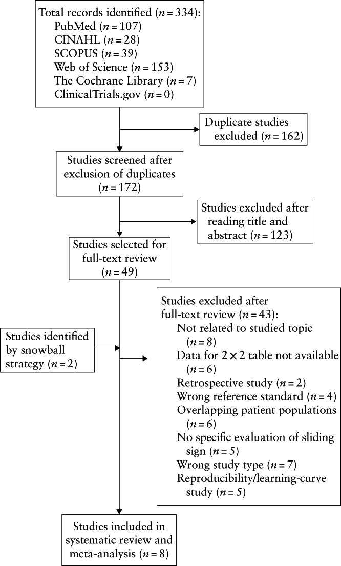
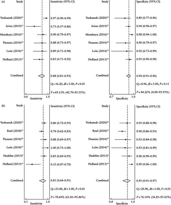
Results: A total of 334 citations were identified. Eight studies were included in the analysis, resulting in 938 and 963 patients available for analysis of the diagnostic accuracy of the sliding sign for pouch of Douglas obliteration and bowel involvement, respectively. The mean prevalence of pouch of Douglas obliteration was 37% and the mean prevalence of bowel involvement was 23%. The pooled estimated sensitivity, specificity, positive likelihood ratio, negative likelihood ratio and diagnostic odds ratio of the sliding sign on TVS for detecting pouch of Douglas obliteration were 88% (95% CI, 81-93%), 94% (95% CI, 91-96%), 15.3 (95% CI, 10.2-22.9), 0.12 (95% CI, 0.07-0.21) and 123 (95% CI, 62-244), respectively. The heterogeneity was moderate for sensitivity and low for specificity for detecting pouch of Douglas obliteration. The pooled estimated sensitivity, specificity, positive likelihood ratio, negative likelihood ratio and diagnostic odds ratio of the sliding sign on TVS for detecting bowel involvement were 81% (95% CI, 64-91%), 95% (95% CI, 91-97%), 16.0 (95% CI, 9.0-28.6), 0.20 (95% CI, 0.10-0.40) and 81 (95% CI, 34-191), respectively. The heterogeneity for the meta-analysis of diagnostic accuracy for bowel involvement was high.
Conclusion: The sliding sign on TVS has good diagnostic performance for predicting pouch of Douglas obliteration and bowel involvement in women with suspected endometriosis. © 2022 The Authors. Ultrasound in Obstetrics & Gynecology published by John Wiley & Sons Ltd on behalf of International Society of Ultrasound in Obstetrics and Gynecology.
Precisión diagnóstica del signo deslizante para detectar la obliteración del fondo de saco de Douglas y la afectación intestinal en mujeres con sospecha de endometriosis: revisión sistemática y metaanálisis
Objetivo: El objetivo de esta revisión sistemática y metaanálisis fue evaluar la precisión diagnóstica del signo deslizante en la ecografía transvaginal (ETV) para detectar la obliteración del fondo de saco de Douglas y la afectación intestinal en pacientes con sospecha de endometriosis, utilizando la laparoscopia como estándar de referencia.
Métodos: Se realizó una búsqueda de estudios que hubieran evaluado la función del signo deslizante en la valoración de la obliteración del fondo de saco de Douglas y/o la afectación intestinal utilizando la laparoscopia como estándar de referencia, publicados desde enero de 2000 hasta octubre de 2021 en las bases de datos PubMed/MEDLINE, Web of Science, CINAHL, The Cochrane Library, ClinicalTrials.gov y SCOPUS. Para evaluar la calidad de los estudios se utilizó la herramienta de Evaluación de Calidad de los Estudios de Precisión Diagnóstica‐2 (QUADAS‐2, por sus siglas en inglés) Los análisis se realizaron mediante los comandos MIDAS y METANDI de STATA.
Resultados: Se identificaron un total de 334 citas. En el análisis se incluyeron ocho estudios, lo que dio como resultado 938 y 963 pacientes disponibles para el análisis de la precisión diagnóstica del signo deslizante para la obliteración del fondo de saco de Douglas y la afectación intestinal, respectivamente. La prevalencia media de la obliteración del fondo de saco de Douglas fue del 37% y la prevalencia media de la afectación intestinal fue del 23%. La estimación combinada de la sensibilidad, especificidad, cociente de verosimilitud positivo, cociente de verosimilitud negativo y razón de momios del diagnóstico del signo deslizante en la ETV para detectar la obliteración del fondo de saco de Douglas fue del 88% (IC 95%, 81–93%), 94% (IC 95%, 91–96%), 15,3 (IC 95%, 10,2–22,9), 0,12 (IC 95%, 0,07–0,21) y 123 (IC 95%, 62–244), respectivamente. La heterogeneidad fue moderada en cuanto a la sensibilidad y baja en cuanto a la especificidad para detectar la obliteración del fondo de saco de Douglas. La estimación combinada de la sensibilidad, especificidad, cociente de verosimilitud positivo, cociente de verosimilitud negativo y razón de momios del diagnóstico del signo deslizante en la ETV para detectar la afectación intestinal fue del 81% (IC 95%, 64–91%), 95% (IC 95%, 91–97%), 16,0 (IC 95%, 9,0–28,6), 0,20 (IC 95%, 0,10–0,40) y 81 (IC 95%, 34–191), respectivamente. La heterogeneidad del metaanálisis de la precisión diagnóstica de la afectación intestinal fue alta.
Conclusiones: El signo deslizante en la ETV tiene un buen rendimiento diagnóstico para predecir la obliteración del fondo de saco de Douglas y la afectación intestinal en mujeres con sospecha de endometriosis.
滑动征检测疑似子宫内膜异位症女性道格拉斯小袋闭塞和肠道受累的诊断准确性:系统评价和荟萃分析
目的: 本系统评价和荟萃分析的目的是 以腹腔镜作为参考标准用于评估经阴道超声 (TVS) 滑动征在检测疑似道格拉斯小袋闭塞和肠道受累并伴有子宫内膜异位患者中的诊断准确性。
方法: 使用2000 年 1 月至 2021 年 10 月出版的《腹腔镜参考标准》,在 PubMed/MEDLINE、Web of Science、CINAHL、Cochrane 图书馆、ClinicalTrials.gov 和 SCOPUS 数据库中搜索滑动征在评估道格拉斯小袋闭塞和/或肠道受累中的作用的研究 。使用诊断准确性研究的质量评价工具‐2(Quality Assessment of Diagnostic Accuracy Studies‐2)来评估这些研究的质量。在STATA 中使用MIDAS 和METANDI 命令进行分析。
结果: 共识别出 334 次引用。共有八项研究被纳入分析,分别有 938 名和 963 名患者可用于分析滑动征对道格拉斯小袋闭塞和肠道受累的诊断准确性。道格拉斯小袋闭塞的平均患病率为 37%,肠道受累的平均患病率为 23%。经阴道超声(TVS )滑动征检测道格拉斯闭塞袋的综合估计敏感性、特异性、阳性似然比、阴性似然比和诊断优势比分别为 88%(95% CI,81‐93%)、94%(95% CI , 91–96%), 15.3 (95% CI, 10.2–22.9), 0.12 (95% CI, 0.07–0.21) 和 123 (95% CI, 62–244)。检测道格拉斯小袋闭塞敏感性中的异质性为中等,特异性低。经阴道超声(TVS )滑动征检测肠道受累的汇总估计敏感性、特异性、阳性似然比、阴性似然比和诊断优势比分别为 81%(95% CI,64‐91%)、95%(95% CI,91 –97%)、16.0 (95%CI, 9.0–28.6)、0.20 (95%CI, 0.10–0.40) 和 81 (95% CI, 34–191)。肠道受累诊断准确性的荟萃分析异质性很高。
结论: 经阴道超声( TVS) 滑动征对预测疑似患有道格拉斯小袋闭塞和肠道受累的子宫内膜异位女性具有良好的诊断性能。
Keywords: bowel; endometriosis; pouch of Douglas; sliding sign; sonography.
© 2022 The Authors. Ultrasound in Obstetrics & Gynecology published by John Wiley & Sons Ltd on behalf of International Society of Ultrasound in Obstetrics and Gynecology.
Figures
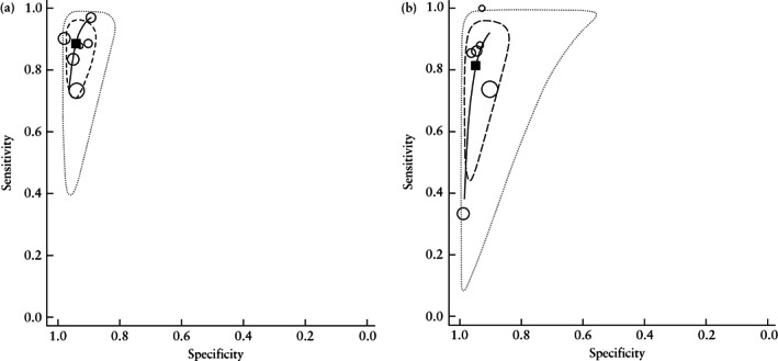
 ) showing performance of the sliding sign on transvaginal ultrasound in the detection of pouch of Douglas obliteration (a) and bowel involvement (b) in women with suspected endometriosis.
  , Study estimate;
  , summary point;
  , 95% confidence region;
  , 95% prediction region.
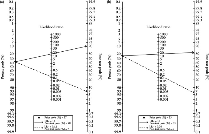
 ) and positive (
 ) sliding sign on transvaginal ultrasound in women with suspected endometriosis. LR−, negative likelihood ratio; LR+, positive likelihood ratio; prob, probability.
Similar articles
-
The prediction of pouch of Douglas obliteration using offline analysis of the transvaginal ultrasound 'sliding sign' technique: inter- and intra-observer reproducibility.Hum Reprod. 2013 May;28(5):1237-46. doi: 10.1093/humrep/det044. Epub 2013 Mar 12. Hum Reprod. 2013. PMID: 23482338
-
Transvaginal ultrasound for detecting parametrial involvement in suspected deep pelvic endometriosis: updated meta-analysis.Ultrasound Obstet Gynecol. 2025 Aug 23. doi: 10.1002/uog.70004. Online ahead of print. Ultrasound Obstet Gynecol. 2025. PMID: 40848275 Review.
-
Can we improve the prediction of pouch of Douglas obliteration in women with suspected endometriosis using ultrasound-based models? A multicenter prospective observational study.Acta Obstet Gynecol Scand. 2015 Dec;94(12):1297-306. doi: 10.1111/aogs.12779. Epub 2015 Oct 23. Acta Obstet Gynecol Scand. 2015. PMID: 26399692
-
Transvaginal ultrasound vs magnetic resonance imaging for diagnosing deep infiltrating endometriosis: systematic review and meta-analysis.Ultrasound Obstet Gynecol. 2018 May;51(5):586-595. doi: 10.1002/uog.18961. Ultrasound Obstet Gynecol. 2018. PMID: 29154402
-
Three-dimensional transvaginal ultrasound vs magnetic resonance imaging for preoperative staging of deep myometrial and cervical invasion in patients with endometrial cancer: systematic review and meta-analysis.Ultrasound Obstet Gynecol. 2022 Nov;60(5):604-611. doi: 10.1002/uog.24967. Ultrasound Obstet Gynecol. 2022. PMID: 35656849 Free PMC article.
Cited by
-
Transvaginal Ultrasound for the Diagnosis of Endometriosis: Current Practices and Barriers in Australian Sonographers.Australas J Ultrasound Med. 2025 May 22;28(2):e70003. doi: 10.1002/ajum.70003. eCollection 2025 May. Australas J Ultrasound Med. 2025. PMID: 40415951
-
Advances in Imaging for Assessing Pelvic Endometriosis.Diagnostics (Basel). 2022 Nov 26;12(12):2960. doi: 10.3390/diagnostics12122960. Diagnostics (Basel). 2022. PMID: 36552967 Free PMC article. Review.
-
Endometriosis imaging in the community setting: implementation of the SRU consensus recommendations on routine pelvic US.Abdom Radiol (NY). 2025 Mar 27. doi: 10.1007/s00261-025-04906-y. Online ahead of print. Abdom Radiol (NY). 2025. PMID: 40146311 Review.
-
Endometriosis MDC: role of the radiologist.Abdom Radiol (NY). 2025 Apr 4. doi: 10.1007/s00261-025-04920-0. Online ahead of print. Abdom Radiol (NY). 2025. PMID: 40183801 Review.
References
-
- Piessens S, Edwards A. Sonographic Evaluation for Endometriosis in Routine Pelvic Ultrasound. J Minim Invasive Gynecol 2020; 27: 265–266. - PubMed
-
- Ghai V, Jan H, Shakir F, Haines P, Kent A. Diagnostic delay for superficial and deep endometriosis in the United Kingdom. J Obstet Gynaecol 2020; 40: 83–89. - PubMed
-
- Piketty M, Chopin N, Dousset B, Millischer‐Bellaische AE, Roseau G, Leconte M, Borghese B, Chapron C. Preoperative work‐up for patients with deeply infiltrating endometriosis: transvaginal ultrasonography must definitely be the first‐line imaging examination. Hum Reprod 2009; 24: 602–607. - PubMed
-
- Holland TK, Hoo WL, Mavrelos D, Saridogan E, Cutner A, Jurkovic D. Reproducibility of assessment of severity of pelvic endometriosis using transvaginal ultrasound. Ultrasound Obstet Gynecol 2013; 41: 210–215. - PubMed
