. 2022:2444:243-269.
doi: 10.1007/978-1-0716-2063-2_15.

Purification and Characterization of Human DNA Ligase IIIα Complexes After Expression in Insect Cells

Affiliations

Purification and Characterization of Human DNA Ligase IIIα Complexes After Expression in Insect Cells

Ishtiaque Rashid et al. Methods Mol Biol. 2022.

Abstract

With improvements in biophysical approaches, there is growing interest in characterizing large, flexible multi-protein complexes. The use of recombinant baculoviruses to express heterologous genes in cultured insect cells has advantages for the expression of human protein complexes because of the ease of co-expressing multiple proteins in insect cells and the presence of a conserved post-translational machinery that introduces many of the same modifications found in human cells. Here we describe the preparation of recombinant baculoviruses expressing DNA ligase IIIα, XRCC1, and TDP1, their subsequent co-expression in cultured insect cells, the purification of complexes containing DNA ligase IIIα from insect cell lysates, and their characterization by multi-angle light scattering linked to size exclusion chromatography and negative stain electron microscopy.

Keywords: Affinity chromatography; Bacmid; Baculovirus; Insect cells; Ion exchange chromatography; Multiple angle light scattering; Negative stain electron microscopy; Size exclusion chromatography.


Figures

Fig. 1
Fig. 1
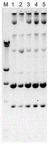
Analysis of recombinant bacmid DNA by 0.5% agarose gel electrophoresis at 20 V for 18–20 h. Bacmid DNA is isolated from five clones per construct (lanes 1–5). Lambda DNA HindIII digest containing 23.1, 9.4, 6.5, 4.3, 2.3, 2.0, and 0.5 kb fragments is used as a DNA size marker (lane M). The signature intact bacmid DNA band is right above the 23.1 kb fragment as indicated by an arrow
Fig. 2
Fig. 2
Small-scale affinity purification of LigIII-XRCC1-TDP1 trimeric complex (left side of gel) and LigIII-TDP1 dimeric complex (right side of gel) from 25 mL of co-infected Sf9 cells using Ni-NTA and anti-Flag M2 columns, respectively. Approximately 10% of first (E1) and second (E2) eluates and 20% of leftover beads (B) were analyzed by 7.5% Tris-Glycine SDS-PAGE with Coomassie staining. Protein molecular weight standards (M) from the top of the gel are 250, 150, 100, 75, and 50 kDa
Fig. 3
Fig. 3
Purification of LigIIIα/TDP1 complex using a nickel column. PL and FT represent pre-load and flow through, respectively. Proteins in 100, 300, and 600 mM imidazole eluates were detected by Coomassie staining after separation by SDS-PAGE
Fig. 4
Fig. 4
Purification of LigIIIα/XRCC1 complex using a dsDNA cellulose column. PL and FT represent pre-load and flow through, respectively. Every other fraction from the peak was analyzed by SDS-PAGE to evaluate protein co-elution as an indicator of complex formation
Fig. 5
Fig. 5
Purification of LigIIIα/TDP1 complex using a HiTrap Q column. PL represents pre-load. Fractions from the peak were analyzed by SDS-PAGE to evaluate protein co-elution as an indicator of complex formation
Fig. 6
Fig. 6
BSA injected onto 3.2/30 (a, c) and 10/300 (b, d) Superdex 200 Increase gel filtration columns showing poor and ideal peak separation, respectively. Adequate peak separation is necessary for proper band broadening correction. The ASTRA software tries to align light scattering (red) and refractive index (blue) detectors in poor (c) and ideal (d) peak separation
Fig. 7
Fig. 7
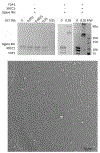
(a) SDS-PAGE gels of LigIIIα complexed with XRCC1, TDP1, or both. In each gel the samples are shown before and after mild chemical crosslinking with GLT. (b) Example negative stain micrograph of LigIIIα crosslinked to TDP1 showing a moderate concentration of particles

References

    1. Abdella R, Talyzina A, Chen S, Inouye CJ, Tjian R, He Y (2021) Structure of the human mediator-bound transcription preinitiation complex. Science 372(6537):52–56 - PMC - PubMed
    1. Chen S, Lee L, Naila T, Fishbain S, Wang A, Tomkinson AE, Lees-Miller SP, He Y (2021) Structural basis of long-range to short-range synapsis in NHEJ. Nature 593(7858):294–298 - PMC - PubMed
    1. Hammel M, Yu Y, Mahaney BL, Cai B, Ye R, Phipps BM, Rambo RP, Hura GL, Pelikan M, So S, Abolfath RM, Chen DJ, Lees-Miller SP, Tainer JA (2010) Ku and DNA-dependent protein kinase dynamic conformations and assembly regulate DNA binding and the initial non-homologous end joining complex. J Biol Chem 285:1414–1423 - PMC - PubMed
    1. Hammel M, Yu Y, Radhakrishnan SK, Chokshi C, Tsai MS, Matsumoto Y, Kuzdovich M, Remesh SG, Fang S, Tomkinson AE, Lees-Miller SP, Tainer JA (2016) An intrinsically disordered APLF links Ku, DNA-PKcs, and XRCC4-DNA ligase IV in an extended flexible non-homologous end joining complex. J Biol Chem 291:26987–27006 - PMC - PubMed
    1. Hammel M, Rashid I, Sverzhinsky A, Pourfarjam Y, Tsai MS, Ellenberger T, Pascal JM, Kim IK, Tainer JA, Tomkinson AE (2021) An atypical BRCT-BRCT interaction with the XRCC1 scaffold protein compacts human DNA ligase IIIalpha within a flexible DNA repair complex. Nucleic Acids Res 49:306–321 - PMC - PubMed

Publication types