. 2022 Mar 14:10:tkac001.
doi: 10.1093/burnst/tkac001. eCollection 2022.

Curcumin-incorporated 3D bioprinting gelatin methacryloyl hydrogel reduces reactive oxygen species-induced adipose-derived stem cell apoptosis and improves implanting survival in diabetic wounds

Affiliations

Curcumin-incorporated 3D bioprinting gelatin methacryloyl hydrogel reduces reactive oxygen species-induced adipose-derived stem cell apoptosis and improves implanting survival in diabetic wounds

Sizhan Xia et al. Burns Trauma. .

Abstract

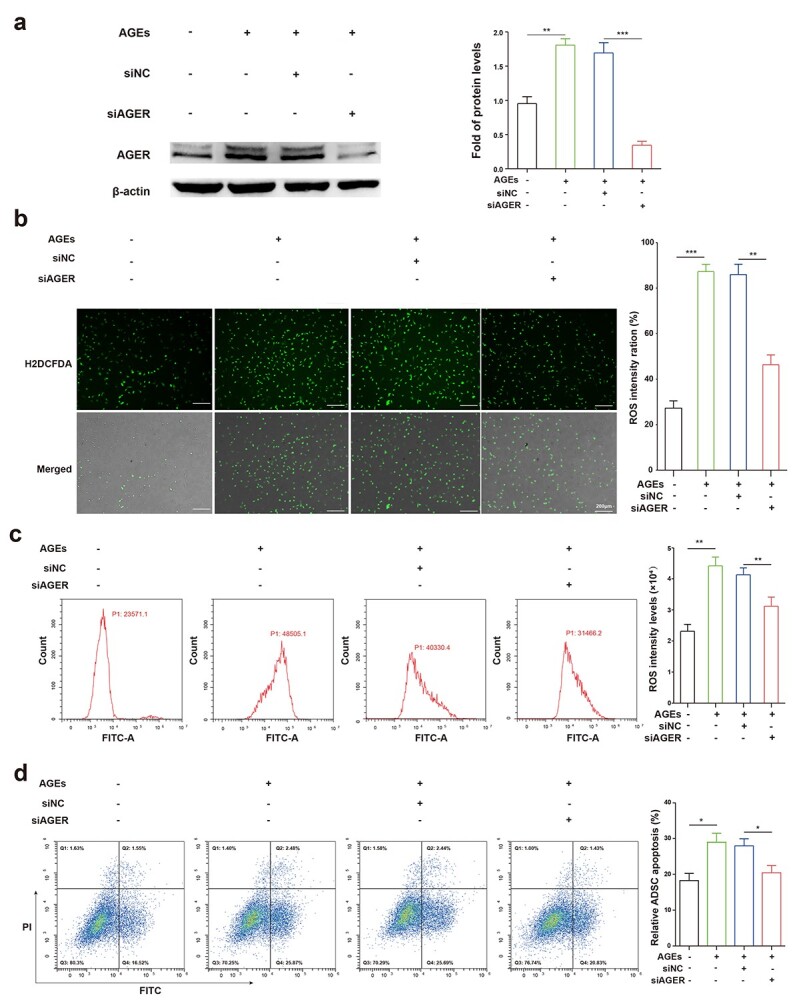
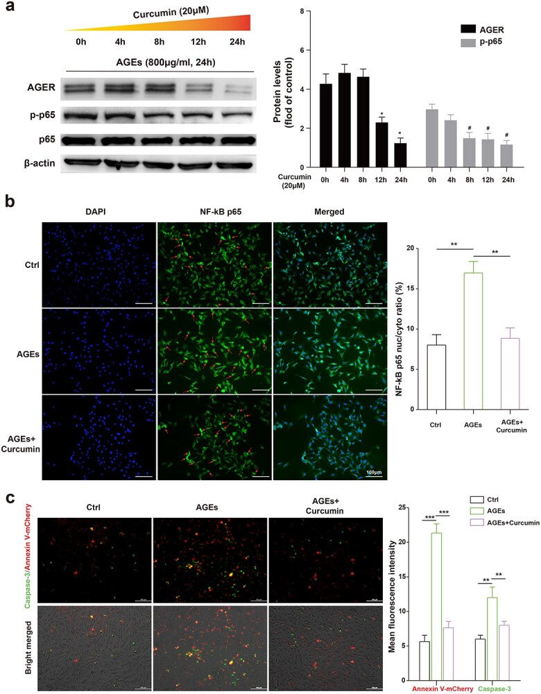
Background: Gelatin methacryloyl (GelMA) hydrogels loaded with stem cells have proved to be an effective clinical treatment for wound healing. Advanced glycation end product (AGE), interacting with its particular receptor (AGER), gives rise to reactive oxygen species (ROS) and apoptosis. Curcumin (Cur) has excellent antioxidant activity and regulates intracellular ROS production and apoptosis. In this study, we developed a Cur-incorporated 3D-printed GelMA to insert into adipose-derived stem cells (ADSCs) and applied it to diabetic wounds.

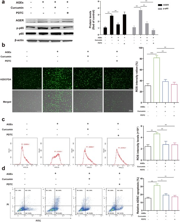
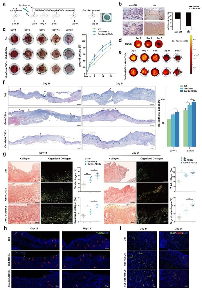
Methods: GelMA hydrogels with Cur were fabricated and their in vitro effects on ADSCs were investigated. We used structural characterization, western blot, ROS and apoptosis assay to evaluate the antioxidant and anti-apoptotic activity, and assessed the wound healing effects to investigate the mechanism underlying regulation of apoptosis by Cur via the AGE/AGER/nuclear factor-κB (NF-κB) p65 pathway.

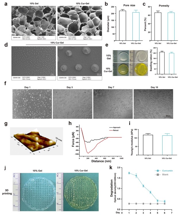
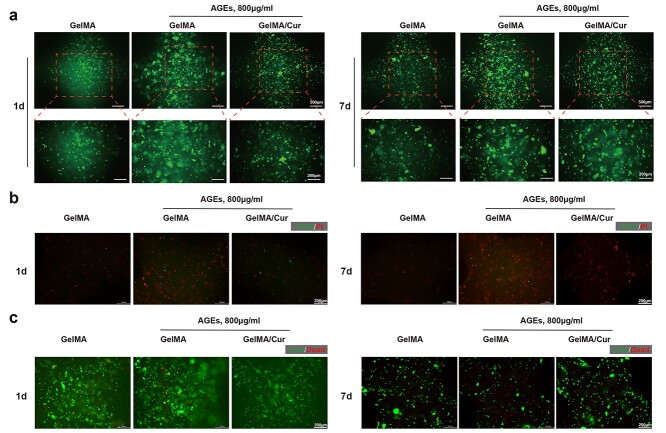
Results: A 10% GelMA scaffold exhibited appropriate mechanical properties and biocompatibility for ADSCs. The circular mesh structure demonstrated printability of 10% GelMA and Cur-GelMA bioinks. The incorporation of Cur into the 10% GelMA hydrogel showed an inhibitory effect on AGEs/AGER/NF-κB p65-induced ROS generation and ADSC apoptosis. Furthermore, Cur-GelMA scaffold promoted cell survival and expedited in vivo diabetic wound healing.

Conclusions: The incorporation of Cur improved the antioxidant activity of 3D-printed GelMA hydrogel and mitigated AGE/AGER/p65 axis-induced ROS and apoptosis in ADSCs. The effects of scaffolds on wound healing suggested that Cur/GelMA-ADSC hydrogel could be an effective biological material for accelerating wound healing.

Keywords: 3D printing; Adipose-derived stem cells; Advanced glycation end products; Curcumin; GelMA; Wound healing.


Figures

Figure 1.
Figure 1.
Structure of GelMA and Cur-GelMA scaffolds. (a) Scanning electron microscopy images of 10% GelMA and 10% Cur-GelMA. (b, c) Quantification of the pore size and porosity of GelMA and Cur-GelMA scaffolds. (d) Scanning electron microscopy images of Cur in GelMA scaffold showing smooth surface morphologies. The mean diameter of particles was 0.99 ± 0.12 μm. (e) Quantification of the swelling ratio of GelMA and Cur-GelMA scaffolds. (f) Representative images of ADSCs on 10% GelMA surfaces. (g–i) Typical atomic force microscopy curve and Young’s modulus of GelMA. (j) Printability test of 10% GelMA and 10% Cur-GelMA hydrogels. (k) Cur degrades in DMEM/F12 with 10% FBS. GelMA Gelatin methacryloyl, Cur curcumin, ADSC adipose-derived stem cell
Figure 2.
Figure 2.
Effects of AGEs on ROS production, and the apoptosis and cell viability of ADSCs on 3D printing scaffolds. (a) Intracellular ROS levels were visualized with a fluorescence microscope. (b) ADSCs were stained with V-FITC/PI and visualized by fluorescence microscope. (c) Cell viability (live/dead fluorescence images) of ADSCs encapsulated in the 10% 3D printing GelMA scaffolds. AGE Advanced glycation end-product, ROS reactive oxygen species, ADSC adipose-derived stem cell, GelMA gelatin methacryloyl
Figure 3.
Figure 3.
Effects of the AGE/AGER axis on ROS production and ADSC apoptosis. (a) Left: western blot shows AGER expression after transfection with siAGER; siRNA served as a negative control for siAGER. Right: quantitative analysis comparing AGER expression levels between the groups. (b) ROS production was tested by fluorescence microscope and the total cell number was observed by bright field microscopy to calculate the percentage. (c) Flow cytometry showing ROS levels in ADSCs. (d) FITC/PI stained ADSCs analyzed by flow cytometry. Data are shown as means ± SD. Statistical analysis: *p<0.05, **p<0.01 and *p<0.05. AGE Advanced glycation end product, AGER AGE receptor, ADSC adipose-derived stem cell, ROS reactive oxygen species
Figure 4.
Figure 4.
Cur suppresses AGEs/AGER axis-mediated ROS and ADSC apoptosis. (a) ADSCs pre-treated with AGEs were mixed with Cur or the p65 inhibitor PDTC (30 μM). Left: western blot demonstrates protein expression levels of AGER and phosphorylated and total p65. Right: comparison of AGER and p-p65 expression levels between the study groups. (b) ROS level was tested by fluorescence microscopy and total cell number was observed by bright field microscopy to calculate the percentage. (c) Flow cytometry showing ROS levels in ADSCs. (d) FITC/PI stained ADSCs analyzed by flow cytometry. Data are shown as means ± SD. Statistical analysis: *p<0.05, **p<0.01 and *p<0.001. AGE Advanced glycation end product, AGER AGE receptor, ROS reactive oxygen species, ADSC adipose-derived stem cell, Cur curcumin, PDTC pyrrolidinedithiocarbamate
Figure 5.
Figure 5.
Role of NF-κB p65 signal in the inhibitory effect of Cur on ADSCs. (a) Left: time course analysis of AGER and phosphorylated and total p65 protein expression levels in ADSCs pre-treated with AGEs (800 μg/mL; 24 h) treated with Cur (20 μM). Right: comparison of AGER and p-p65 expression levels between the study groups. (b) ADSCs pre-treated with AGEs (800 μg/mL; 24 h) with or without Cur (20 μM; 24 h). Translocation of p65 from the cytoplasm to the nucleus was quantified by cell immunofluorescence staining (red arrowheads). (c) V-mCherry/caspase-3 stained ADSCs analyzed by flow cytometry. Relative fluorescence intensity was used to calculate cell apoptosis and caspase-3 expression level. Data are shown as means ± SD. Statistical analysis: *p<0.05, **p<0.01 and *p<0.001. (*pvs 0 h of AGER, #pvs 0 h of p-p65). NF-κB nuclear factor-κB, Cur curcumin, ADSC adipose-derived stem cell, AGE advanced glycation end product, AGER AGE receptor
Figure 6.
Figure 6.
Cur-GelMA scaffold enhances ADSC survival and accelerates diabetic wound healing. (a) Above: schematic diagram of in vivo wound healing of nude mice. Below: schematic representation of 3D bioprinted GelMA scaffolds and in vivo transplantation. (b) Representative immunohistochemistry of AGEs in nude mice and streptozocin-induced nude mice skin tissue. Quantification of AGEs+ cells (%) analyzed using ImageJ software. (c) Left: gross appearance of the skin wounds after administration of GelMA, GelMA-ADSCs or Cur-GelMA-ADSCs. Excisional wound-splinting assay demonstrating improved wound closure with Cur-GelMA-ADSCs compared to the other scaffolds. Right: wound healing rate of each group. (d) IVIS was used to detect DiR fluorescence after DiR labelling of ADSCs transplanted on the surface of excisional wounds on days 0, 3 and 7. (e). IVIS was used to detect DiR fluorescence after DiR labelling of ADSCs in 3D printing scaffolds transplanted in excisional wounds on days 0, 3, 7, 14 and 21. The color scale is shown on the right side. (f) Left: representative H&E-stained sections on days 14 and 21 after wound creation. Pink arrows point to the epithelial tongues and green arrows to the GelMA of interest. Right: Percentage of re-epithelialization between the evaluated groups. (g) Left: representative SR-stained section on days 14 and 21 after wound creation. Right: the percentage of total collagen and organized collagen between the groups. (h) TUNEL fluorescence staining of wounds from the three groups on days 14 and 21 showed apoptosis (red arrowheads). (i) Representative immunofluorescence images on days 14 and 21 after wound creation showing the presence of CD31+ and α-SMA+ vessels. Data are shown as means ± SD. Statistical analysis: **p<0.05, and ***p<0.01. (*pvs Gel-ADSCs, #pvs Gel.) Gel-MA gelatin methacryloyl, ADSC adipose-derived stem cell, Cur curcumin, AGE advanced glycation end product, DiR 1,1′-dioctadecyl-3,3,3′,3′-tetramethylindotricarbocyanine, IVIS in vivo imaging system, H&E hematoxylin and eosin, SR swelling ratio

Similar articles

Cited by

References

    1. Frykberg RG, Banks J. Challenges in the treatment of chronic wounds. Adv Wound Care (New Rochelle). 2015;4:560–82. - PMC - PubMed
    1. Rathmann W, Giani G. Global prevalence of diabetes: estimates for the year 2000 and projections for 2030. Diabetes Care. 2004;27:2568–9author reply 9. - PubMed
    1. Game F. Treatment strategies for neuroischaemic diabetic foot ulcers. Lancet Diabetes Endocrinol. 2018;6:159–60. - PubMed
    1. Eke G, Mangir N, Hasirci N, MacNeil S, Hasirci V. Development of a UV crosslinked biodegradable hydrogel containing adipose derived stem cells to promote vascularization for skin wounds and tissue engineering. Biomaterials. 2017;129:188–98. - PubMed
    1. Wu Y, Liang T, Hu Y, Jiang S, Luo Y, Liu C, et al. . 3D bioprinting of integral ADSCs-NO hydrogel scaffolds to promote severe burn wound healing. Regen Biomater. 2021;8:rbab014. - PMC - PubMed