

Clinical Scenario and Imaging with Illustrations of Giant Cell Tumor of Bone: A Retrospective Analysis
- PMID: 35291246
- PMCID: PMC8889423
- DOI: 10.22038/ABJS.2021.50922.2522
Clinical Scenario and Imaging with Illustrations of Giant Cell Tumor of Bone: A Retrospective Analysis
Abstract
Background: The giant cell tumour of the bone has a spectrum of clinical-radiological presentation. This study aims to describe this varied presentation in our institution.
Methods: This retrospective study was conducted on twenty-nine pathologically labelled cases of giant cell tumours of the bone. The medical records for their clinical presentation and diagnostic imaging studies were studied and evaluated.
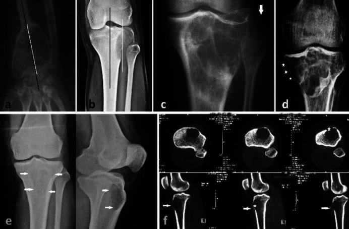
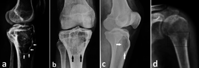
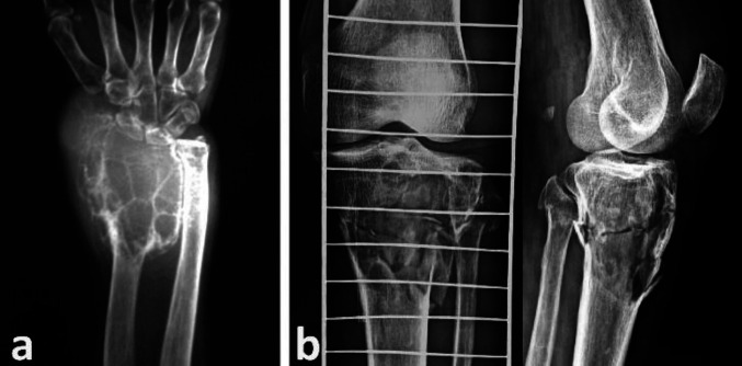
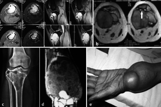
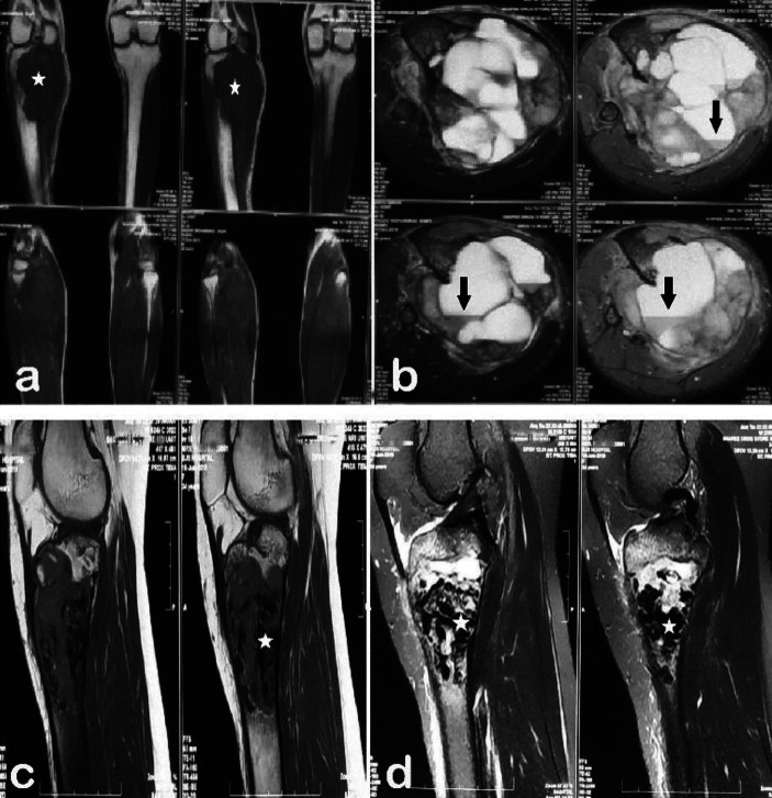
Results: Mean age of the patients at presentation was 35.3±12.9 years. Pain, local swelling and restricted joint function were seen in 93 %, 58.6 % and 52 % patients, respectively. The cortical breach was seen in 15 (51.7 %) and 22 (75.9 %) lesions on plain radiographs and CT images, respectively. 14(48.3 %) cases had soft tissue invasion on MRI at presentation. 26 (89.7 %) lesions were located within 1 cm from the articular cartilage. The solid tumour component was hypo to iso-intense in signal intensity in 27 (93.1 %) lesions in T1 weighted and 21 (72.4 %) in T2 weighted images. 14 (48.3 %) had hyperintense cystic areas, and fluid-fluid levels, suggestive of aneurysmal bone cysts, were seen in 4 (13.8 %) cases on T2 weighted images. Hypo-echoic nodular areas in solid tumour component, suggestive of hemosiderin deposits, were present in 3 (10.3 %) lesions on T1 and T2 weighted images.
Conclusion: The tumour classically presents as an epiphysial-metaphyseal, eccentric, expansile, lytic lesion in a skeletally mature patient. The MRI picture is variable and the surgeon should have a sound knowledge of these variations to obtain a biopsy sample from a proper site of the lesion and to avoid misdiagnosis especially of a primary ABC.
Keywords: ABC; Fluid-Fluid; Giant-Cell; Hemosiderin; Soap-Bubble.
Conflict of interest statement
We do not have any conflict of interest in publication of this research work.
Figures


References
-
- Murphey MD, Nomikos GC, Flemming DJ, Gannon FH, Temple HT, Kransdorf MJ. Imaging of giant cell tumor and giant cell reparative granuloma of bone: radiologic-pathologic correlation. Radiographics. 2001;21(5):1283–309. - PubMed
-
- Forsyth RG, De Boeck G, Bekaert S, De Meyer T, Taminiau AH, Uyttendaele D, et al. Telomere biology in giant cell tumour of bone. The Journal of Pathology: A Journal of the Pathological Society of Great Britain and Ireland. 2008;214(5):555–63. - PubMed
-
- McGrath PJ. Giant-cell tumour of bone: an analysis of fifty-two cases. J Bone Joint Surg Br. 1972;54(2):216–29. - PubMed
LinkOut - more resources
Full Text Sources