Large granular lymphocytic leukemia: a brief review
- PMID: 35291253
- PMCID: PMC8918699
Large granular lymphocytic leukemia: a brief review
Abstract
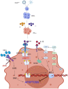
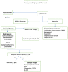
LGL leukemia is a rare chronic lymphoproliferative disorder of cytotoxic lymphocytes which can be immunophenotypically either T cell or NK cell-derived. According to the World Health Organization classification, it can be divided into three subtypes: chronic T-cell leukemia and chronic natural killer cell lymphocytosis, and aggressive natural killer cell LGL leukemia. Clonal proliferation of large granular lymphocytes can be because of stimulation of various molecular pathways namely JAK-STAT3 pathway, FAS/FAS-L pathway, RAS-RAF-1-MEK1-ERK pathway, PI3K/AKT pathway, NF-KB pathway, and Sphingolipid Rheostat pathways. The most common clinical features presenting with this leukemia are neutropenia, anemia, thrombocytopenia. This leukemia is also associated with various autoimmune conditions. It usually has an indolent course except for the aggressive NK cell LGL leukemia. The cause of death in the indolent cases was mostly due to infectious complications related to the neutropenia associated with the disease. The rarity of the disease coupled with the availability of only a handful of clinical trials has been a hindrance to the development of a specific treatment. Most of the cases are managed with immunomodulators. The advances in the knowledge of molecular pathways associated with the disease have brought few targeted therapies into the limelight. We discuss here the evolution, epidemiology, demographic profile, pathophysiology, differential diagnosis, the available treatment options along with the survival and prognostic variables which may help us in better understanding and better management of the disease and hopefully, paving the way for a targeted clinical approach.
Keywords: LGL leukemia; NK cell; T cell.
AJBR Copyright © 2022.
Conflict of interest statement
None.
Figures


Similar articles
-
Latest Advances in the Diagnosis and Treatment of Large Granular Lymphocytic Leukemia.Am Soc Clin Oncol Educ Book. 2018 May 23;38:616-625. doi: 10.1200/EDBK_200689. Am Soc Clin Oncol Educ Book. 2018. PMID: 30231346 Review.
-
Molecular Features and Diagnostic Challenges in Alpha/Beta T-Cell Large Granular Lymphocyte Leukemia.Int J Mol Sci. 2022 Nov 2;23(21):13392. doi: 10.3390/ijms232113392. Int J Mol Sci. 2022. PMID: 36362180 Free PMC article.
-
LGL leukemia: from pathogenesis to treatment.Blood. 2017 Mar 2;129(9):1082-1094. doi: 10.1182/blood-2016-08-692590. Epub 2017 Jan 23. Blood. 2017. PMID: 28115367 Review.
-
The complex relationship between large granular lymphocyte leukemia and rheumatic disease.Expert Rev Clin Immunol. 2024 Mar;20(3):291-303. doi: 10.1080/1744666X.2023.2292758. Epub 2023 Dec 18. Expert Rev Clin Immunol. 2024. PMID: 38105745 Review.
-
Large Granular Lymphocytic Leukemia: Clinical Features, Molecular Pathogenesis, Diagnosis and Treatment.Cancers (Basel). 2024 Mar 27;16(7):1307. doi: 10.3390/cancers16071307. Cancers (Basel). 2024. PMID: 38610985 Free PMC article. Review.
Cited by
-
Rare Hematologic Malignancies and Pre-Leukemic Entities in Children and Adolescents Young Adults.Cancers (Basel). 2024 Feb 29;16(5):997. doi: 10.3390/cancers16050997. Cancers (Basel). 2024. PMID: 38473358 Free PMC article. Review.
-
T-PLL Presenting with an Indolent Course.Case Rep Hematol. 2024 Jul 24;2024:7310135. doi: 10.1155/2024/7310135. eCollection 2024. Case Rep Hematol. 2024. PMID: 39104431 Free PMC article.
-
Sepsis Unveils T-cell Large Granular Lymphocytic Leukemia in the Setting of End-Stage Renal Disease: A Rare Hematologic Malignancy.Cureus. 2024 Mar 1;16(3):e55325. doi: 10.7759/cureus.55325. eCollection 2024 Mar. Cureus. 2024. PMID: 38562340 Free PMC article.
-
Plurality Over Parsimony: When Two Diagnoses Are More Likely Than One.J Gen Intern Med. 2024 May;39(7):1257-1263. doi: 10.1007/s11606-023-08585-7. Epub 2024 Feb 26. J Gen Intern Med. 2024. PMID: 38409513 Free PMC article. No abstract available.
-
A case report of T-LGL leukemia-associated pure red cell aplasia harboring STAT3, TNFAIP3, and KMT2D mutation.Transl Cancer Res. 2023 Apr 28;12(4):1054-1059. doi: 10.21037/tcr-23-326. Epub 2023 Apr 10. Transl Cancer Res. 2023. PMID: 37180665 Free PMC article.
References
-
- Brouet JC, Sasportes M, Flandrin G, Preud’Homme JL, Seligmann M. Chronic lymphocytic leukaemia of T-cell origin immunological and clinical evaluation in eleven patients. Lancet. 1975;306:890–893. - PubMed
-
- McKenna RW, Parkin J, Kersey JH, Gajl-Peczalska KJ, Peterson L, Brunning RD. Chronic lymphoproliterative disorder with unusual clinical, morphologic, ultrastructural and membrane surface marker characteristics. Am J Med. 1977;62:588–596. - PubMed
-
- Agnarsson BA, Loughran TP Jr, Starkebaum G, Kadin ME. The pathology of large granular lymphocyte leukemia. Hum Pathol. 1989;20:643–651. - PubMed
-
- Lougharn TP. Clonal diseases of large granular lymphocytes. Blood. 1993;82:12–14. - PubMed
Publication types
LinkOut - more resources
Full Text Sources
Research Materials
Miscellaneous
