. 2022 Jan;10(4):518-526.
doi: 10.52547/rbmb.10.4.518.

A Comparative Study to Visualize PtdIns(4,5) P2 and PtdIns(3,4,5) P3 in MDA-MB-231 Breast Cancer Cell Line

Affiliations

A Comparative Study to Visualize PtdIns(4,5) P2 and PtdIns(3,4,5) P3 in MDA-MB-231 Breast Cancer Cell Line

Dhurgham Al-Fahad et al. Rep Biochem Mol Biol. 2022 Jan.

Abstract

Background: Phosphatidylinositol 3,4,5-trisphosphate [PtdIns(3,4,5) P3) and Phosphatidylinositol 4,5-trisphosphate (PtdIns(4,5) P2] form an insignificant amount of phospholipids but play important roles in controlling membrane-bound signalling. Little attention has been given to visualize and monitor changes or differences in the local generation of PtdIns(4,5) P2 and PtdIns(3,4,5) P3 in the cell membranes of MDA-MB-231 breast cancer cell lines.

Methods: PLCδ1-PH-GFP and Btk-PH-GFP were used as biosensors to detected PtdIns(4,5) P2 and PtdIns(3,4,5)P3 respectively. These biosensors and antibodies were transfected, immuostained and then visualized by confocal microscopy on different cell surfaces.

Results: Our results showed that PLCδ1-PH-GFP/mCherry was localized at the cell membrane, while Btk-PH-GFP/mCherry was sometimes localized at the cell membrane but there was also a large amount of fluorescence present in the cytosol and nucleus. Our results also showed that the cells that expressed low levels of Btk-PH-GFP the fluorescence was predominantly localised to the cell membrane. While the cells that expressed high levels of Btk-PH-GFP the fluorescence was localization in the cytosol and cell membrane. Our results demonstrated that both anti-PtdIns(4,5)P2 and anti-PtdIns(3,4,5)P3 antibodies were localized everywhere in cell.

Conclusion: Our results suggest that PLCδ1-PH-GFP and Btk-PH-GFP/mCherry have more specificity, reliability, suitability and accuracy than antibodies in binding with and detecting PtdIns(4,5)P2 and PtdIns(3,4,5)P3 and in studying the molecular dynamics of phospholipids in live and fixed cells.

Keywords: Antibodies; Biosensors; MDA-MB-231; Phosphatidylinositol.


Figures
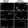
Fig. 1
Fig. 1
visualisation of PtdIns(4,5)P2 and PtdIns(3,4,5)P3on the variety of surfaces in fixed cells: Confocal images of MDA-MB-231 cells expressing the PLCδ1-GFP or mCherry and Btk-PH-GFP were used as biosensors to identify PtdIns(4,5)P2 and PtdIns(3,4,5)P3lipids in the plasma membrane of MDA-MB-321 cells respectively. MDA-MB-321 cells were seeded on the variety of surfaces (gelatine 0.2%, fibronectin 20 µg/ml and collagen 2mg/ml). Then, cells were transfected with PLCδ1-GFP or mCherry and Btk-PH-GFP or mCherry after stimulating PtdIns(3,4,5) P3productionwith EGF (100 ng/ml) for 5 minutes. Then, cells were fixed with paraformaldehyde to visualise PtdIns(4,5) P2 and PtdIns(3,4,5) P3. The green arrow indicates the localisation of PtdIns(4,5)P2 and PtdIns(3,4,5)P3 on the cell membrane and the red arrow indicates the localisation of PtdIns(3,4,5)P3in the cytosol. Three independent experiments were performed, representative pictures are shown.
Fig. 2
Fig. 2
Effect of Btk-PH-GFP overexpression in the cell. Confocal images of MDA-MB-231 cells that were transfected with Btk-PH-GFP for 24-hour, A) At low expression levels, the peak of intensity in the cell membrane was higher than in the cytosol. B) At high expression levels, the peak of intensity in the cell membrane and cytosol was similar. The white arrow indicates the localisation of PtdIns(3,4,5) P3in the cell membrane and the red arrow indicates the localisation of PtdIns(3,4,5) P3in the cytosol. The red line across cell refers to the measurement of intensity in the cell membrane and cytosol of the cell. Data are a representative of three independent experiments in which 10 cells were measured.
Fig. 3
Fig. 3
Quantification of PtdIns(4,5) P2 localisation. A) MDA-MB-231 cells were transfected with PLCδ1-PH-GFP and fixed with paraformaldehyde. Quantification of intensity in the cell membrane and cytosol of the cell. The peak of intensity was only in the cell membrane. B) MDA-MB-231 cells were stained with anti-PtdIns(4,5) P2antibody and fixed with paraformaldehyde. Quantification of intensity in the cell membrane and cytosol of the cell. The peak of intensity in the cell membrane and cytosol was similar. Data are representative of three independent experiments in which 10 cells were measured.
Fig. 4
Fig. 4
Quantification of PtdIns(3,4,5)P3 localization. A) MDA-MB-231 cells were transfected with Btk-PH-GFP and fixed with paraformaldehyde. Quantification of the intensity in the cell membrane and cytosol. The peak of intensity in the cell membrane was higher than in the cytosol. B) MDA-MB-231 cells were stained with anti-PtdIns(3,4,5) P3antibody and then fixed with paraformaldehyde. Quantification of the intensity in the cell membrane and cytosol of the cell. The peak of intensity in the cell membrane and cytosol was similar. Data are representative of three independent experiments in which 10 cells were measured.

References

    1. Mashaghi S, Jadidi T, Koenderink G, Mashaghi A. Lipid Nanotechnology. Int J Mol Sci. 2013;14(2):4242–4282. - PMC - PubMed
    1. Wang X, Hills LB, Huang YH. Lipid and Protein Co-Regulation of PI3K Effectors Akt and Itk in Lymphocytes. Front Immunol. 2015;6:117. - PMC - PubMed
    1. Lemmon MA. Pleckstrin Homology (PH) domains and phosphoinositides. Biochem Soc Symp. 2007;74:81–93. - PMC - PubMed
    1. Alfahad D, Alharethi S, Alharbi B, Mawlood K, Dash P. PtdIns(4,5)P2 and PtdIns(3,4,5)P3 dynamics during focal adhesions assembly and disassembly in a cancer cell line. Turk J Biol. 2020;44(6):381–92. - PMC - PubMed
    1. Santos CR, Schulze A. Lipid metabolism in cancer. The FEBS journal. 2012;279(15):2610–23. - PubMed

LinkOut - more resources