Review
. 2022 Feb 14;22(1):e7.
doi: 10.4110/in.2022.22.e7. eCollection 2022 Feb.

Immunopathology and Immunotherapy of Inflammatory Skin Diseases

Affiliations
Review

Immunopathology and Immunotherapy of Inflammatory Skin Diseases

Ahreum Song et al. Immune Netw. .

Abstract

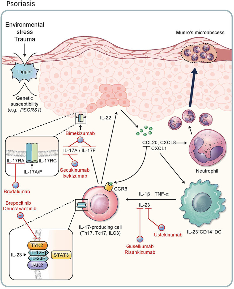
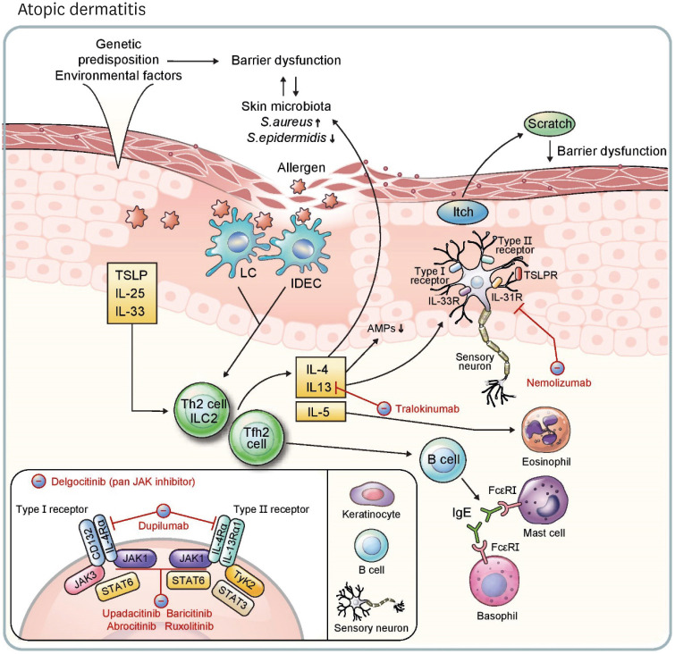
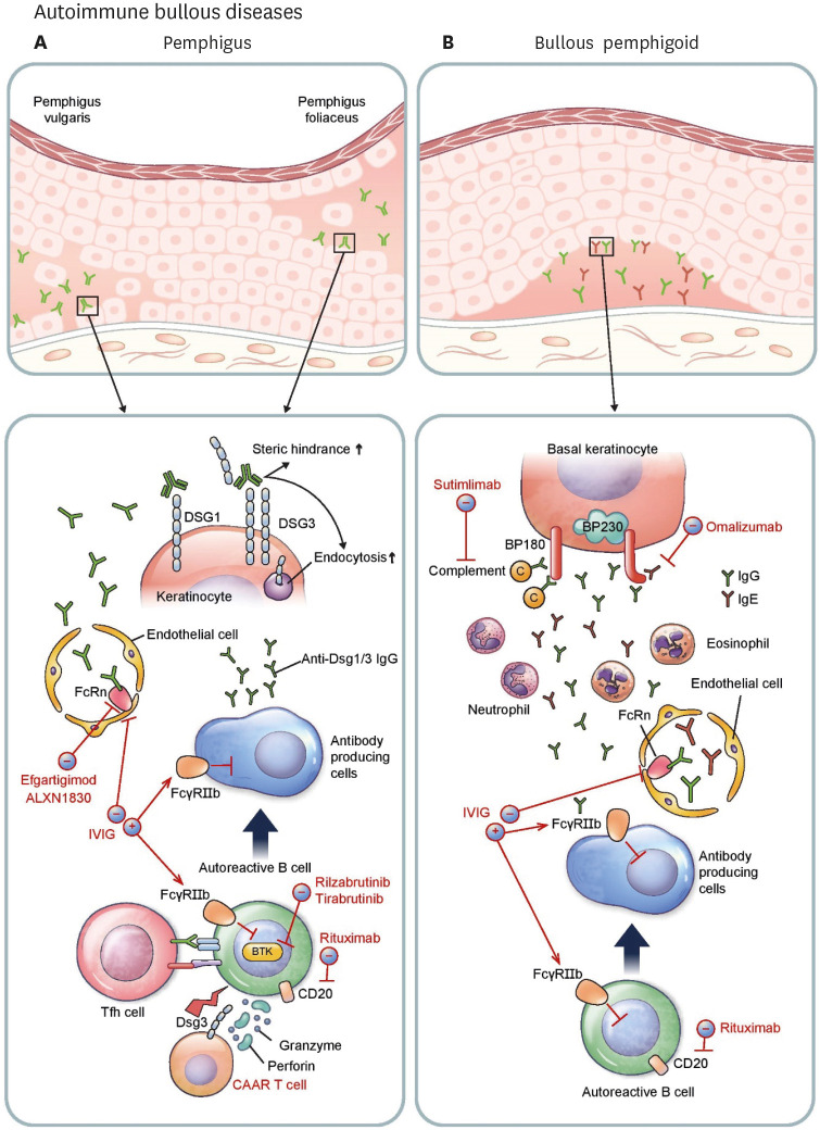
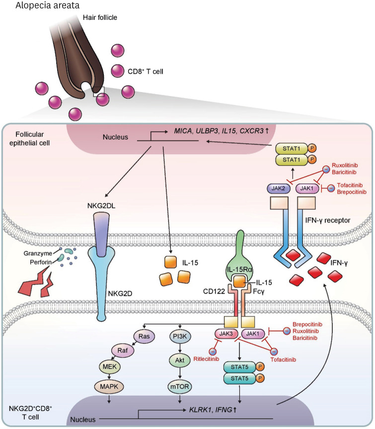
Recently, there have been impressive advancements in understanding of the immune mechanisms underlying cutaneous inflammatory diseases. To understand these diseases on a deeper level and clarify the therapeutic targets more precisely, numerous studies including in vitro experiments, animal models, and clinical trials have been conducted. This has resulted in a paradigm shift from non-specific suppression of the immune system to selective, targeted immunotherapies. These approaches target the molecular pathways and cytokines responsible for generating inflammatory conditions and reinforcing feedback mechanisms to aggravate inflammation. Among the numerous types of skin inflammation, psoriasis and atopic dermatitis (AD) are common chronic cutaneous inflammatory diseases. Psoriasis is a IL-17-mediated disease driven by IL-23, while AD is predominantly mediated by Th2 immunity. Autoimmune bullous diseases are autoantibody-mediated blistering disorders, including pemphigus and bullous pemphigoid. Alopecia areata is an organ-specific autoimmune disease mediated by CD8+ T-cells that targets hair follicles. This review will give an updated, comprehensive summary of the pathophysiology and immune mechanisms of inflammatory skin diseases. Moreover, the therapeutic potential of current and upcoming immunotherapies will be discussed.

Keywords: Alopecia areata; Atopic dermatitis; Biologics; Immunopathology; Pemphigoid, bullous; Pemphigus; Psoriasis.


Conflict of interest statement

Conflicts of Interest: The authors declare no potential conflicts of interest.

Figures

Figure 1
Figure 1. Pathophysiology of and immunotherapeutic agents for psoriasis. External environmental stress and trauma is a possible trigger of psoriasis, especially in individuals with genetic susceptibilities (e.g., PSORS1). This leads to the release of CCL20, CXCL8, and CXCL1, stimulating CCR6+IL-17+ lymphocytes, neutrophils, and IL-23+CD14+ DCs, respectively. Release of IL-17A and IL-17F via IL-17-producing cells (Th17, Tc17, and ILC3) results in the activation of the IL-17RA/IL-17RC complex in keratinocytes, further feeding the inflammatory response. Production of IL-22 by ILC3 accelerates hyperproliferation of keratinocytes. DCs induce cytokines like IL-1β, TNF-α, and IL-23. IL-17-producing cells are stimulated by IL-23 through the JAK2/TYK2-STAT3 pathway. Ustekinumab targets the p40 subunit shared by IL-12 and IL-23, and guselkumab and risankizumab inhibit the p19 subunit of IL-23. Secukinumab and ixekizumab are inhibitors of IL-17A. Bimekizumab is a monoclonal antibody targeting IL-17A and IL-17F. Brodalumab is an inhibitor of IL-17RA. With respect to JAK signaling, deucravacitinib acts to inhibit TYK2, and brepocitinib inhibits TYK2 and JAK1 simultaneously.
Figure 2
Figure 2. Pathophysiology of and immunotherapeutic agents for atopic dermatitis. Genetic predisposition and environmental factors induce barrier dysfunction. Further, change of surface microbiome diversity, especially decrease of S. epidermidis and increase of S. aureus, enhances barrier dysfunction and increases vulnerability of skin epidermis to external allergens. TSLP, IL-25, and IL-33 released from keratinocytes promote a type 2 response through ILC2, Th2 cells, and Tfh2 cells, which are induced from activated skin LCs and IDECs. IL-4, IL-13, and IL-5 are released from these lymphocytes. IL-4 and IL-13 activate type I (IL-4Rα/CD132) and type II (IL-4Rα/IL-13Rα1) receptors on B cells, keratinocytes, and sensory neurons, resulting in activation of JAK-STAT pathways. Tfh2 cells induce IgE+ B cells, stimulating mast cells and basophils via FcεRI. AMPs (e.g., human β-defensin 3) are decreased in keratinocytes in response to IL-4 and IL-13. Neuronal itch is induced by IL-4, IL-13, IL-31, IL-33 and TSLP, and the itch-scratch cycle is intensified through the process. Type 2 cytokines also aggravate the imbalance of skin surface microbiota, and IL-5 recruits eosinophils, continuing the vicious circle. Dupilumab inhibits type I and II receptors by blocking IL-4Rα. Tralokinumab neutralizes IL-13 and nemolizumab inhibits IL-31R. Upadacitinib and abrocitinib are oral, selective JAK1 inhibitors, and baricitinib is an oral JAK1 and JAK2 inhibitor. Topical agents ruxolitinib (a JAK1 and JAK2 inhibitor) and delgocitinib (a pan-JAK inhibitor) are approved treatments for atopic dermatitis.
Figure 3
Figure 3. Pathophysiology and immunotherapeutic agents of autoimmune bullous diseases. (A) In pemphigus, Tfh cells activate autoreactive B cells, which differentiate into antibody-producing cells generating pathogenic anti-Dsg1/3 autoantibodies. Autoantibodies circulate through vessels and relocate to the epidermal intercellular space. FcRn expressed on endothelial cells lengthens the half-life of IgG autoantibodies. Binding of autoantibodies to Dsg1/3 induces steric hindrance and enhances endocytosis, leading to loss of cell-to-cell adhesion of keratinocytes. Rituximab depletes autoreactive B cells by targeting cell surface antigen CD20. Rilzabrutinib and tirabrutinib are small molecule drugs that bind BTK in B cells and inhibit aberrant B-cell receptor signaling. Dsg3 CAAR T cells specifically target and destroy Dsg3-specific B cells. ALXN1830 and efgartigimod shorten the half-life of antibodies by blocking FcRn. Intravenous immunoglobulin (IVIG) inhibits antibody-producing cells by activating the FcγRIIb inhibitory receptor and enhances the degradation of autoantibodies by saturating FcRn. (B) In BP, autoreactive B cells switch to become antibody-producing cells resulting in production of pathogenic anti-BP180/BP230 autoantibodies which migrate to dermo-epidermal junction. IgG and IgE autoantibodies against BP180 promote complement activation leading to infiltration of granulocytes such as neutrophils and eosinophils into the dermis. Release of various proteases plays an important role in creating clefts and in blister formation. Along with pemphigus, rituximab depletes CD20+ B cells. IVIG saturates FcRn reducing the life span of autoantibodies, and suppresses antibody-producing cells by activating FcγRIIb. Omalizumab, a monoclonal antibody against IgE, can decrease the level of pathogenic IgE autoantibodies. Sutimlimab decreases complement activation at the dermo-epidermal junction.
Figure 4
Figure 4. Pathophysiology and immunotherapeutic agents of alopecia areata. In alopecia areata, NKG2D+CD8+ T cells infiltrate into the dermis and relocate to the hair follicle bulb. IL-15 is an important cytokine for pathogenesis of this disease through activation and proliferation of NKG2D+CD8+ T cells. The IL-15 receptor complex is composed of IL-15Rα expressed on follicular epithelial cells and CD122 and Fcγ on T cells, and trans-activates CD8+ T cells through multiple pathways including Ras-Raf-MEK-MAPK, PI3K-Akt-mTOR, and JAK1/3-STAT5 signaling. These pathways upregulate expression of KLRK1, encoding NKG2D, and IFNG. IFN-γ binds to the IFN-γ receptor on follicular epithelial cells and triggers JAK1/2-STAT1 signaling, thereby upregulating expression of MICA, ULBP3, IL15, and CXCR3. Ruxolitinib and baricitinib inhibit JAK1 and JAK2, and brepocitinib inhibits JAK1. Tofacitinib is a JAK1 and JAK3 inhibitor, and ritlecitinib is a selective JAK3 inhibitor.

References

    1. Greb JE, Goldminz AM, Elder JT, Lebwohl MG, Gladman DD, Wu JJ, Mehta NN, Finlay AY, Gottlieb AB. Psoriasis. Nat Rev Dis Primers. 2016;2:16082. - PubMed
    1. Alinaghi F, Calov M, Kristensen LE, Gladman DD, Coates LC, Jullien D, Gottlieb AB, Gisondi P, Wu JJ, Thyssen JP, et al. Prevalence of psoriatic arthritis in patients with psoriasis: a systematic review and meta-analysis of observational and clinical studies. J Am Acad Dermatol. 2019;80:251–265.e219. - PubMed
    1. Heydendael VM, Spuls PI, Opmeer BC, de Borgie CA, Reitsma JB, Goldschmidt WF, Bossuyt PM, Bos JD, de Rie MA. Methotrexate versus cyclosporine in moderate-to-severe chronic plaque psoriasis. N Engl J Med. 2003;349:658–665. - PubMed
    1. Lee E, Trepicchio WL, Oestreicher JL, Pittman D, Wang F, Chamian F, Dhodapkar M, Krueger JG. Increased expression of interleukin 23 p19 and p40 in lesional skin of patients with psoriasis vulgaris. J Exp Med. 2004;199:125–130. - PMC - PubMed
    1. Nair RP, Duffin KC, Helms C, Ding J, Stuart PE, Goldgar D, Gudjonsson JE, Li Y, Tejasvi T, Feng BJ, et al. Genome-wide scan reveals association of psoriasis with IL-23 and NF-kappaB pathways. Nat Genet. 2009;41:199–204. - PMC - PubMed