

doi: 10.1186/s13024-022-00529-9.
The route of SARS-CoV-2 to brain infection: have we been barking up the wrong tree?
Affiliations
- PMID: 35292068
- PMCID: PMC8922388
- DOI: 10.1186/s13024-022-00529-9
Item in Clipboard
The route of SARS-CoV-2 to brain infection: have we been barking up the wrong tree?
Mol Neurodegener.
.
Abstract
This letter draws attention to recent work supporting the notion that the SARS-CoV-2 virus may use the nervus terminalis rather than the olfactory nerve as a shortcut route from the nasal cavity to infect the brain.
Keywords: ACE2; Brain infection; COVID-19; Nervus terminalis; Olfactory system; Omicron; SARS-CoV-2; TMPRSS2.
© 2022. The Author(s).
Conflict of interest statement
The authors declare no competing interests.
Figures
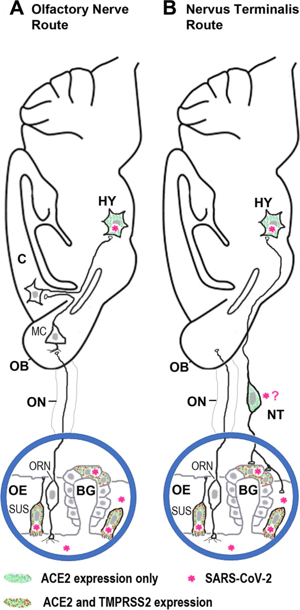
Schematic illustration of two routes how SARS-CoV-2 or the virus’ cleaved S1 subunit of the spike protein may travel from the nose to the brain. A. Route along the olfactory nerve (ON). Olfactory receptor neurons (ORNs) and most mitral cells (MCs) in the olfactory bulb (OB) do not express the obligatory viral entry protein, ACE2 (angiotensin-converting enzyme 2), are rarely or not at all infected by the virus, and the ON and the OB are not always infected when SARS-CoV-2 is found in the brain [3, 9]. Support cells in the olfactory epithelium express ACE2 and the surface protease TMPRSS2, and these cells (sustentacular cells, SUS) become infected with SARS-CoV-2 [3, 7, 8]. C, cerebral cortex; Hy, hypothalamus. B. Route along the nervus terminalis (NT). This cranial nerve connects the olfactory epithelium, and in particular Bowman gland (BG) cells, directly with nuclei beyond the olfactory bulb (OB), including the hypothalamus [11, 12]. Support cells (SUS) and BG cells express ACE2 and TMPRSS2 and are known to become infected by SARS-CoV-2 [3, 7, 8]. Nervus terminalis (NT) neurons also express ACE2, as do neurons in the hypothalamus (HY) which become infected by SARS-CoV-2 [9]. NT neurons and endocrine neurons in the hypothalamus do not express TMPRSS2, but they express neuropilin 1 [9] or cathepsins [12], other proteases that can mediate virus membrane fusion. Whether NT neurons become infected by SARS-CoV-2 or by its cleaved S1 spike protein remains to be determined
References
Publication types
MeSH terms
Grants and funding
LinkOut - more resources
Full Text Sources
Medical
Miscellaneous
