. 2022 Mar:77:103912.
doi: 10.1016/j.ebiom.2022.103912. Epub 2022 Mar 13.

Orally-active, clinically-translatable senolytics restore α-Klotho in mice and humans

Affiliations

Orally-active, clinically-translatable senolytics restore α-Klotho in mice and humans

Yi Zhu et al. EBioMedicine. 2022 Mar.

Abstract

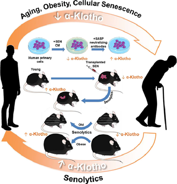
Background: α-Klotho is a geroprotective protein that can attenuate or alleviate deleterious changes with ageing and disease. Declines in α-Klotho play a role in the pathophysiology of multiple diseases and age-related phenotypes. Pre-clinical evidence suggests that boosting α-Klotho holds therapeutic potential. However, readily clinically-translatable, practical strategies for increasing α-Klotho are not at hand. Here, we report that orally-active, clinically-translatable senolytics can increase α-Klotho in mice and humans.

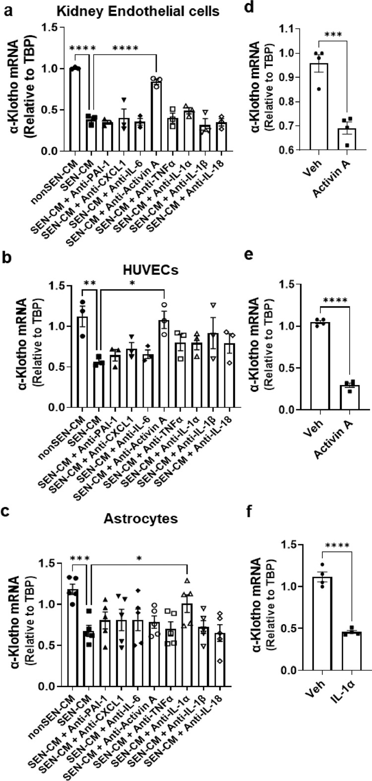
Methods: We examined α-Klotho expression in three different human primary cell types co-cultured with conditioned medium (CM) from senescent or non-senescent cells with or without neutralizing antibodies. We assessed α-Klotho expression in aged, obese, and senescent cell-transplanted mice treated with vehicle or senolytics. We assayed urinary α-Klotho in patients with idiopathic pulmonary fibrosis (IPF) who were treated with the senolytic drug combination, Dasatinib plus Quercetin (D+Q).

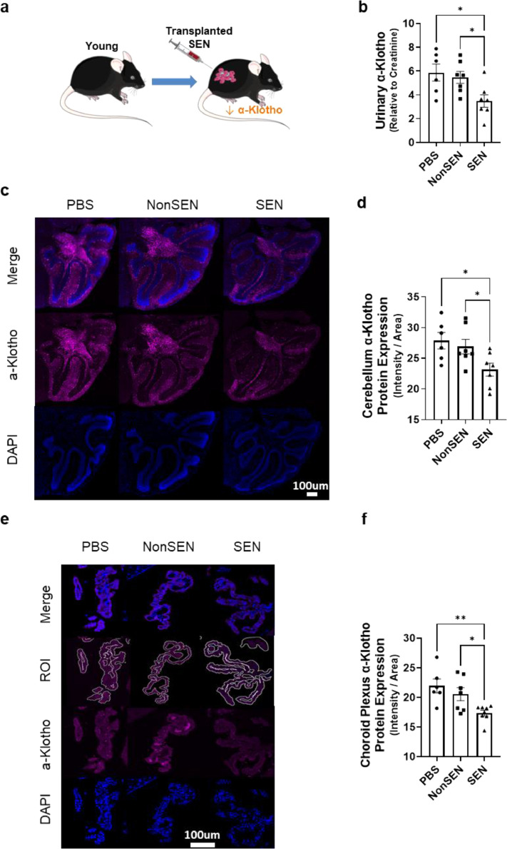
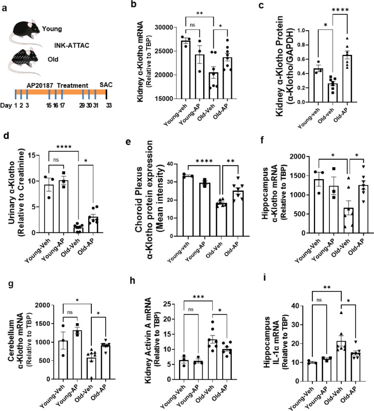
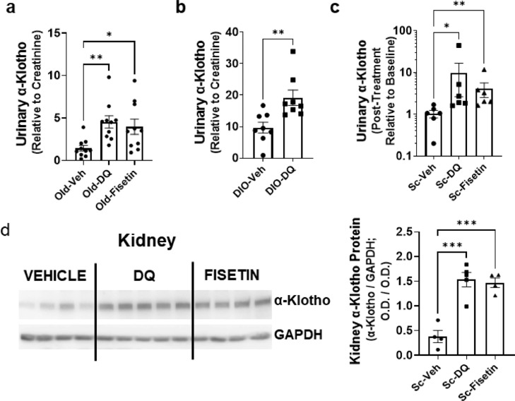
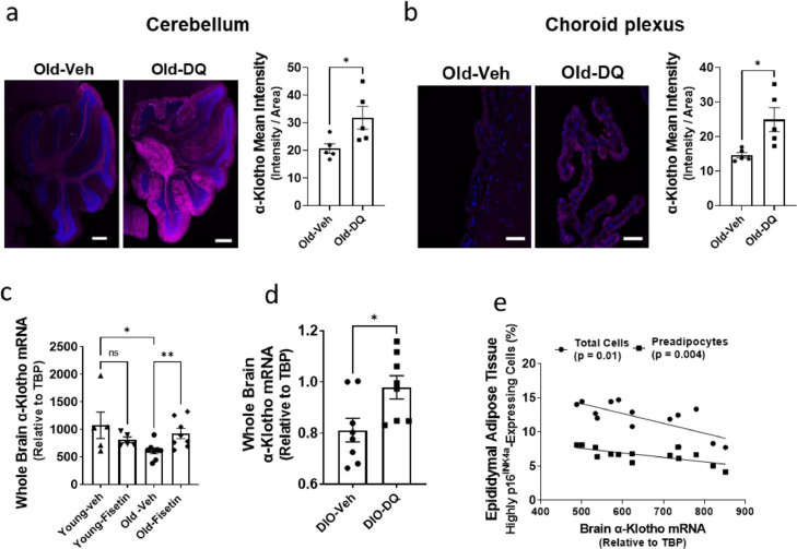
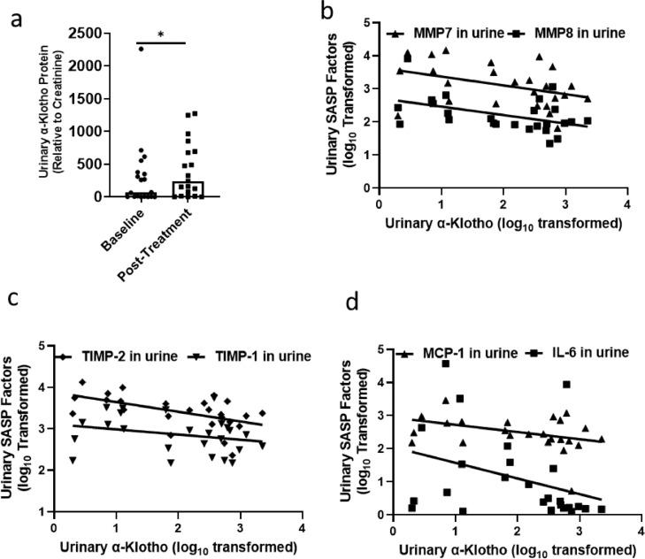
Findings: We found exposure to the senescent cell secretome reduces α-Klotho in multiple nonsenescent human cell types. This was partially prevented by neutralizing antibodies against the senescence-associated secretory phenotype (SASP) factors, activin A and Interleukin 1α (IL-1α). Consistent with senescent cells' being a cause of decreased α-Klotho, transplanting senescent cells into younger mice reduced brain and urine α-Klotho. Selectively removing senescent cells genetically or pharmacologically increased α-Klotho in urine, kidney, and brain of mice with increased senescent cell burden, including naturally-aged, diet-induced obese (DIO), or senescent cell-transplanted mice. D+Q increased α-Klotho in urine of patients with IPF, a disease linked to cellular senescence.

Interpretation: Senescent cells cause reduced α-Klotho, partially due to their production of activin A and IL-1α. Targeting senescent cells boosts α-Klotho in mice and humans. Thus, clearing senescent cells restores α-Klotho, potentially opening a novel, translationally-feasible avenue for developing orally-active small molecule, α-Klotho-enhancing clinical interventions. Furthermore, urinary α-Klotho may prove to be a useful test for following treatments in senolytic clinical trials.

Funding: This work was supported by National Institute of Health grants AG013925 (J.L.K.), AG062413 (J.L.K., S.K.), AG044271 (N.M.), AG013319 (N.M.), and the Translational Geroscience Network (AG061456: J.L.K., T.T., N.M., S.B.K., S.K.), Robert and Arlene Kogod (J.L.K.), the Connor Group (J.L.K.), Robert J. and Theresa W. Ryan (J.L.K.), and the Noaber Foundation (J.L.K.). The previous IPF clinical trial was supported by the Claude D. Pepper Older Americans Independence Centers at WFSM (AG021332: J.N.J., S.B.K.), UTHSCA (AG044271: A.M.N.), and the Translational Geroscience Network.

Keywords: Cellular senescence; Senolytics; α-Klotho.


Conflict of interest statement

Declaration of interests Y.Z., T.T., N.G., T.P., A.K.P., and J.L.K. have a financial interest related to this research. Patents on senolytic drugs are held by Mayo Clinic. This research has been reviewed by the Mayo Clinic Conflict of Interest Review Board and was conducted in compliance with Mayo Clinic Conflict of Interest policies. No conflicts of interest, financial or otherwise, are declared by the other authors.

Figures

Image, graphical abstract
Graphical abstract
Figure1
Figure 1
Senescent cell conditioned medium decreases α-Klotho; blocking particular SASP factors with neutralizing antibodies partially restores α-Klotho. (a-c) Conditioned media (CM) collected from senescent or non-senescent (a) human primary kidney endothelial cells (HKEs; one-way ANOVA: F = 19.10, p < 0.0001, R2 = 0.89; n = 3; ****p < 0.0001); (b) human umbilical vein endothelial cells (HUVECs; one-way ANOVA: F = 3.02, p = 0.02, R2 = 0.58, n = 3; *p = 0.02, **p = 0.01); or (b) human brain astrocytes (HBAs; one-way ANOVA: F = 7.93, p = 0.02, R2 = 0.66; n = 5; ***p < 0.001, *p = 0.03) were applied to non-senescent HKEs, HUVECs, or HBAs, respectively, with or without antibodies against PAI-1, CXCL1, IL-6, activin A, TNF-α, IL-1α, IL-1β, or IL-18 for 48 h. α-Klotho mRNA was assayed by qPCR. Means ± SEM. (d) Primary HKEs (n = 4), (e) HUVECs (n = 4), or (f) HBAs (n = 4) were treated with recombinant activin A or IL-1α. α-Klotho was assayed by qPCR 24 h after treatments. Means ± SEM; unpaired T-tests; ***p < 0.001, ****p < 0.0001.
Figure2
Figure 2
Transplanting senescent cells decreases urinary and brain α-Klotho. (a) 8-month-old male mice were transplanted i.p. with 2 million senescent or non-senescent mouse preadipocytes in 150 µl PBS through a 22-G needle. (b) Urinary α-Klotho was assayed by ELISA and expressed as a function of creatinine (one-way ANOVA: F = 6.45, p = 0.01, R2 = 0.42; n = 7; **p < 0.01, *p = 0.02). Representative images of immunofluorescent (IF) α-Klotho stains of cerebellum (c) and choroid plexus (e) are shown. ROI (Region of Interest, outlined in white) were quantified by ImageJ (d,f) from mice that had been transplanted with senescent or non-senescent cells. Means ± SEM; one-way ANOVA and post hoc Tukey's tests: F = 4.94, p = 0.02, R2 = 0.38. *p = 0.02, *p = 0.03 in (d), F = 7.84; p <0.01, R2 = 0.38, **p < 0.01, *p = 0.01 in (f).
Figure3
Figure 3
Genetic clearance of highly p16Ink4a-expressing cells increases α-Klotho. Young (8-month-old) or old (27-29-month-old) INK-ATTAC male mice were treated with vehicle or AP20187 (n = 3 young+ vehicle; n = 3 young+ AP20187; n = 7 old + vehicle; n = 7 old + AP20187) to dimerize the FKBP-caspase-8 fusion protein expressed in highly p16Ink4a-expressing cells to selectively eliminate p16Ink4a+ cells. AP20187 (10 mg/kg) or vehicle was administered i.p. every 2 weeks for 6 weeks. (a) Schematic view of treatments. (b) Kidney α-Klotho mRNA was assayed by qPCR. One-way ANOVA, F = 4.81, p = 0.01, R2 = 0.46; *p = 0.03, **p < 0.01. (c) α-Klotho protein was assayed relative to GAPDH (Western blots in Supplementary Fig. 7). One-way ANOVA: F = 4.81, p = 0.01, R2 = 0.46; *p < 0.05, ****p < 0.0001. (d) Mouse urine was collected 2 days after the last dose of AP20187. Urinary α-Klotho was assayed by ELISA and expressed as a function of creatinine. One-way ANOVA, F = 61.53, p < 0.00001, R2 = 0.90; *p = 0.04, ****p < 0.0001. Choroid Plexus (e) α-Klotho protein was assayed by immunofluorescence. One-way ANOVA, F = 20.98, R2 = 0.80, **p < 0.01, ****p < 0.0001. Hippocampal (f) and cerebellar (g) α-Klotho mRNA was assayed by qPCR. Activin A mRNA in kidney (h) and IL-1α mRNA in hippocampus (i) were assayed by qPCR. Means ± SEM; one-way ANOVA and post hoc Tukey's tests; F = 3.84, p = 0.038, R2 = 0.40; *p = 0.04 in (f); *p = 0.04, F = 7.87, p < 0.01, R2 = 0.60; *p = 0.03 in (g); F = 10.85; p < 0.001, R2 = 0.64; *p = 0.03, ***p < p < 0.0001 in (h); F = 5.49; p = 0.01, R2 = 0.49; *p < 0.05, **p = 0.01 in (i).
Figure4
Figure 4
Senolytics increase urinary and kidney α-Klotho in old or obese mice. (a) Naturally-aged male mice (28-29-month-old, n = 10 in each group) were treated with vehicle, Dasatinib plus Quercetin (D+Q), or Fisetin. Urinary α-Klotho was measured by ELISA and expressed as a function of creatinine. One-way ANOVA: F = 6.39; p < 0.05, R2 = 0.30; *p = 0.02, **p < 0.01. (b) DIO mice (male, 10-month-old, n = 8) were treated with vehicle or D+Q and urinary α-Klotho was assayed by ELISA and expressed as a function of creatinine. Unpaired T test; **p < 0.01. (c) Six-month-old male mice (n = 6 in each group) were transplanted i.p. with senescent preadipocytes. After 3 months, they were treated with vehicle, D+Q, or Fisetin. Urinary α-Klotho was assayed by ELISA and expressed as a function of creatinine. One-way ANOVA and post hoc Tukey's tests: Kruskal-Wallis statistic=10.19; *p = 0.02, **p < 0.01. (d) Kidney α-Klotho protein was also assayed in the same transplanted mice by Western blotting. One-way ANOVA: F = 24.24; p < 0.001, R2 = 0.83; ***p < 0.001.
Figure5
Figure 5
Senolytics increase brain α-Klotho in old mice. Naturally-aged mice (female, 28-month-old) were treated with vehicle (n = 5) or D+Q (n = 5). Brain α-Klotho was analysed by IF. (a) Representative IF images of α-Klotho expression (red) in the cerebellum are shown. Mean intensities of red florescence in the cerebellum were quantified by ImageJ. Unpaired T-tests; *p = 0.03. (b) Brain choroid plexus α-Klotho (purple), representative IF images. Mean intensities of florescence in the choroid plexus were quantified by ImageJ. Unpaired T-tests; *p = 0.02. (c) Young (female, 6-month-old, n = 5 in vehicle and treated groups) and naturally-aged mice (female, 22-month-old, n = 8 in vehicle and treated groups) were treated with Fisetin or vehicle. α-Klotho in whole brain was assayed by qPCR. One-way ANOVA and post hoc Tukey's tests: Kruskal-Wallis statistic=9.49; *p = 0.02, **p < 0.01. (d) DIO mice (male, 8-9-month-old) were treated with D+Q (n = 8) or vehicle (n = 8). Brain α-Klotho was assayed by qPCR. Mann-Whitney test; *p = 0.03. (e) Correlation between brain α-Klotho mRNA and peripheral p16Ink4a-expressing adipose tissue progenitor cells was quantified by CyTOF. Spearman correlation analysis.
Figure6
Figure 6
Senolytics increase α-Klotho in human urine; urinary SASP factors are inversely related to urinary α-Klotho. Subjects (n = 20) with idiopathic pulmonary fibrosis (IPF) were administered 3 courses of D+Q for 3 sequential days each (total 9 doses over 3 weeks). (a) Urinary α-Klotho and SASP factors were assayed at baseline and 5 days after the last D+Q treatment. Urinary α-Klotho in subjects with IPF was increased (Baseline: 293.49±115.48, Post-treatment: 392.79±95.24, normalized to urinary creatinine) by D+Q. Wilcoxon signed rank tests (two-sided) were used to test differences before and after treatment. *p = 0.04. (b–d) Spearman correlations of urinary α-Klotho and SASP factors are shown. FDR-corrected R2 and p values are in Supplementary Table 2.

References

    1. Maique J., Flores B., Shi M., et al. High phosphate induces and klotho attenuates kidney epithelial senescence and fibrosis. Front Pharmacol. 2020;11:1273. - PMC - PubMed
    1. Kuro OM. The klotho proteins in health and disease. Nat Rev Nephrol. 2019;15(1):27–44. - PubMed
    1. Kuro OM. Klotho and endocrine fibroblast growth factors: Markers of chronic kidney disease progression and cardiovascular complications? Nephrol Dial Transpl. 2019;34(1):15–21. - PubMed
    1. Hu M.C., Shiizaki K., Kuro-o M., Moe OW. Fibroblast growth factor 23 and klotho: Physiology and pathophysiology of an endocrine network of mineral metabolism. Annu Rev Physiol. 2013;75:503–533. - PMC - PubMed
    1. Delcroix V., Mauduit O., Tessier N., et al. The role of the anti-aging protein klotho in igf-1 signaling and reticular calcium leak: impact on the chemosensitivity of dedifferentiated liposarcomas. Cancers. 2018;10(11):439. (Basel) - PMC - PubMed