Review
. 2022 Apr:200:105281.
doi: 10.1016/j.antiviral.2022.105281. Epub 2022 Mar 12.

Global update on the susceptibilities of human influenza viruses to neuraminidase inhibitors and the cap-dependent endonuclease inhibitor baloxavir, 2018-2020

Affiliations
Review

Global update on the susceptibilities of human influenza viruses to neuraminidase inhibitors and the cap-dependent endonuclease inhibitor baloxavir, 2018-2020

Elena A Govorkova et al. Antiviral Res. 2022 Apr.

Abstract

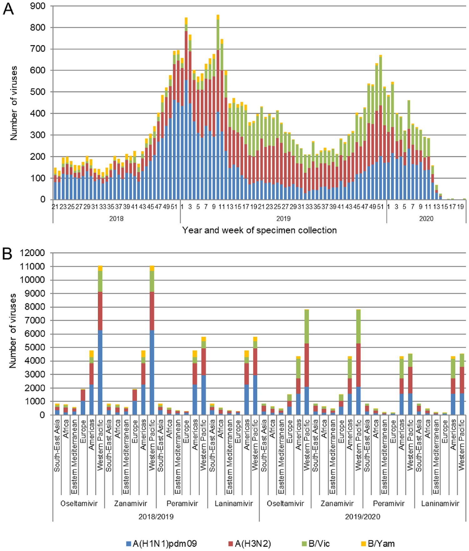
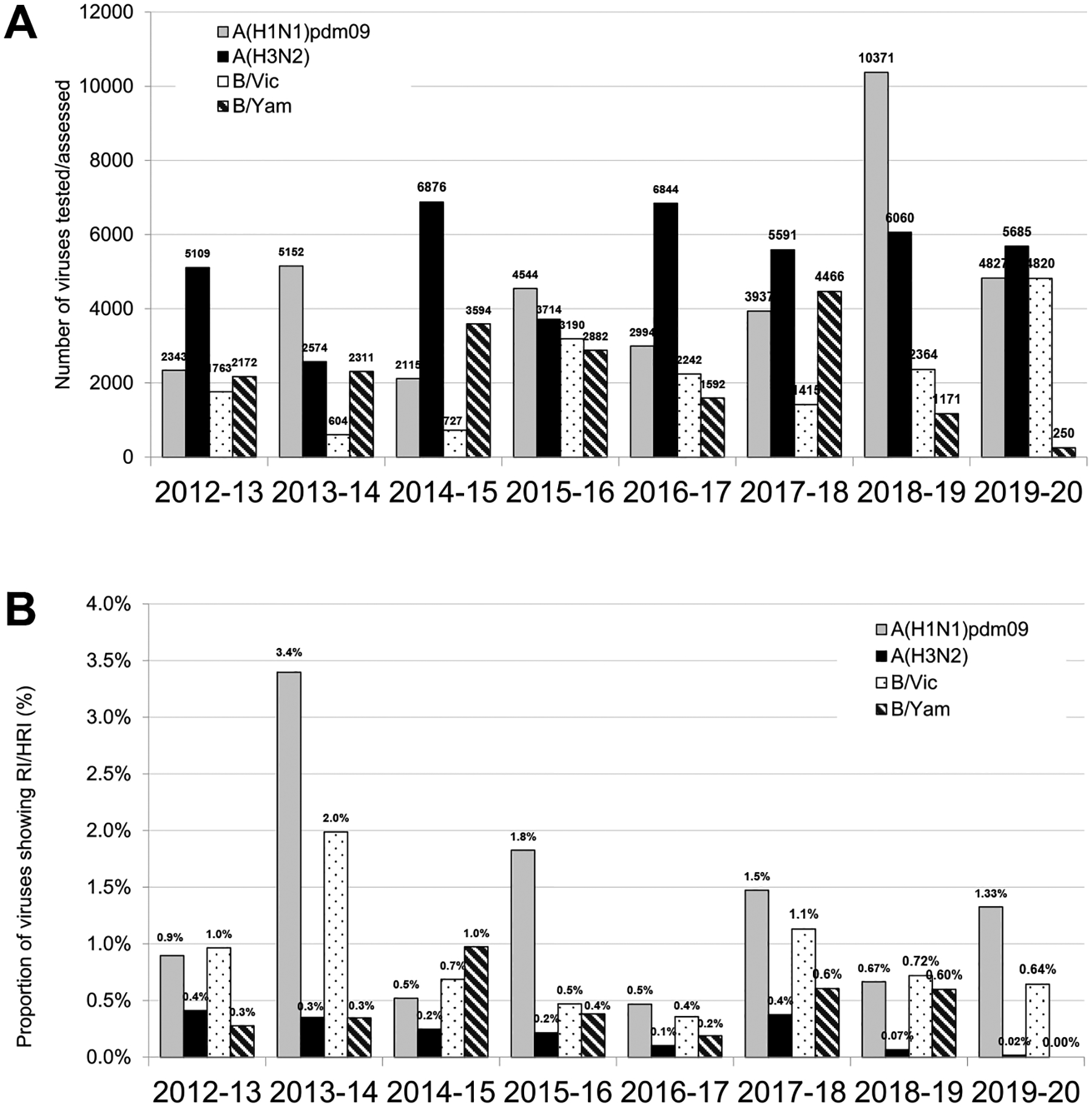
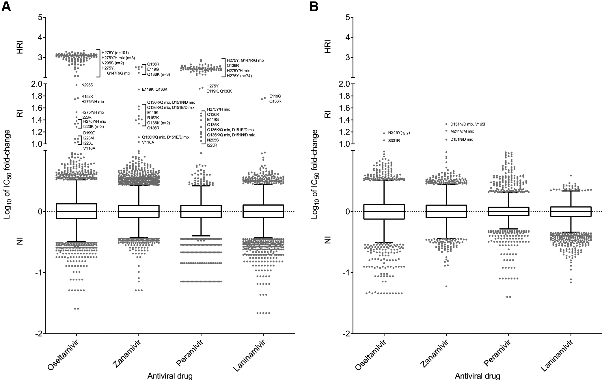
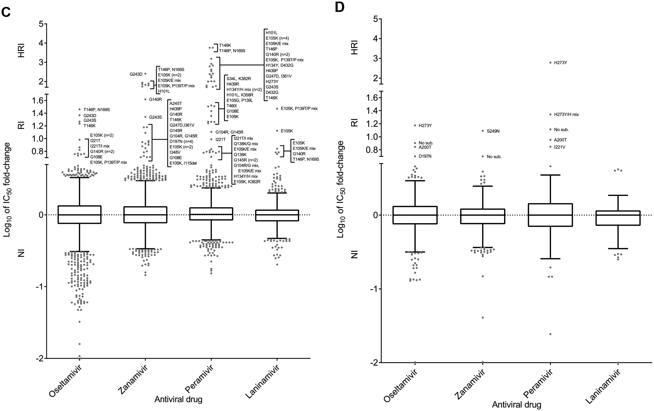
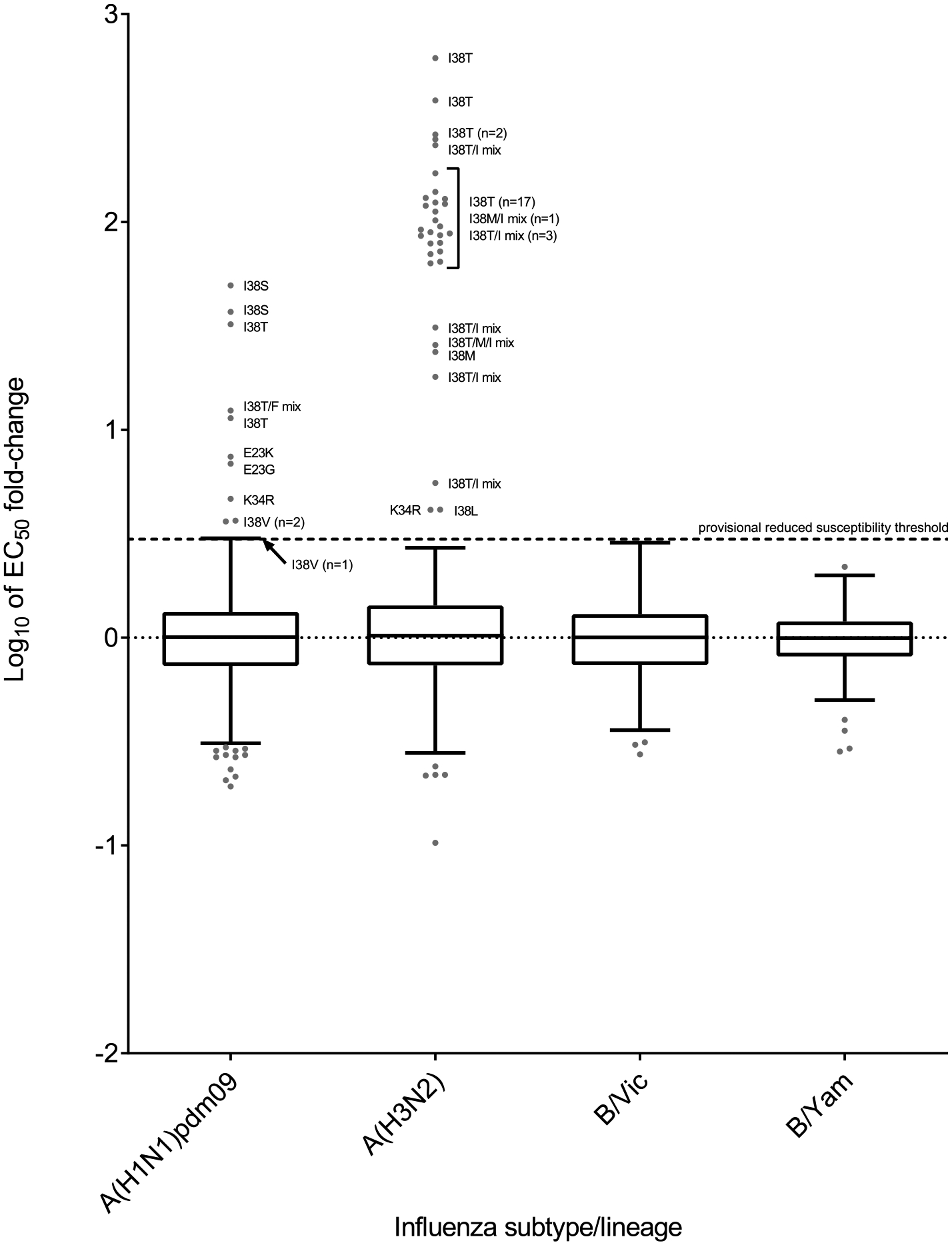
Global analysis of the susceptibility of influenza viruses to neuraminidase (NA) inhibitors (NAIs) and the polymerase acidic (PA) inhibitor (PAI) baloxavir was conducted by five World Health Organization Collaborating Centres for Reference and Research on Influenza during two periods (May 2018-May 2019 and May 2019-May 2020). Combined phenotypic and NA sequence-based analysis revealed that the global frequency of viruses displaying reduced or highly reduced inhibition (RI or HRI) or potential to show RI/HRI by NAIs remained low, 0.5% (165/35045) and 0.6% (159/26010) for the 2018-2019 and 2019-2020 periods, respectively. The most common amino acid substitution was NA-H275Y (N1 numbering) conferring HRI by oseltamivir and peramivir in A(H1N1)pdm09 viruses. Combined phenotypic and PA sequence-based analysis showed that the global frequency of viruses showing reduced susceptibility to baloxavir or carrying substitutions associated with reduced susceptibility was low, 0.5% (72/15906) and 0.1% (18/15692) for the 2018-2019 and 2019-2020 periods, respectively. Most (n = 61) of these viruses had I38→T/F/M/S/L/V PA amino acid substitutions. In Japan, where baloxavir use was highest, the rate was 4.5% (41/919) in the 2018-2019 period and most of the viruses (n = 32) had PA-I38T. Zoonotic viruses isolated from humans (n = 32) in different countries did not contain substitutions in NA associated with NAI RI/HRI phenotypes. One A(H5N6) virus had a dual substitution PA-I38V + PA-E199G, which may reduce susceptibility to baloxavir. Therefore, NAIs and baloxavir remain appropriate choices for the treatment of influenza virus infections, but close monitoring of antiviral susceptibility is warranted.

Keywords: Antiviral; Baloxavir; Influenza; Neuraminidase inhibitor; Polymerase inhibitor; Reduced susceptibility.


Conflict of interest statement

Declaration of interests

The authors declare that they have no known competing financial interests or personal relationships that could have appeared to influence the work reported in this paper.

Figures

Fig. 1.
Fig. 1.
Influenza viruses collected and assessed for NAI susceptibility during 2018–2019 and 2019–2020 periods. For each period viruses were collected between week 21 and week 20 of the following year. (A) Week of specimen collection and virus type/subtype/lineage for specimens assessed in the 2018–2019 and 2019–2020 periods. Typically, week 21 to week 39 of a year covers the Southern Hemisphere influenza season, while week 40 of a year to week 20 of the following year covers the Northern Hemisphere influenza season. (B) The number of viruses assessed for susceptibility to the four NAIs using NA inhibition assays and/or sequence-based analysis, by WHO Region for the 2018–2019 and the 2019–2020 periods.
Fig. 2.
Fig. 2.
Comparison of NAI susceptibility surveillance over eight periods. (A) Number of viruses tested. For the 2012–2018 period testing was reported based on NA inhibition assays only. For the 2018–2020 period results of assessment by NA inhibition assays and/or sequence-based analysis were included. (B) The proportion of viruses showing RI/HRI by NAIs over the 2012–2020 period. Data were compiled from the global studies of viruses isolated during the 2012–2013 (Meijer et al., 2014), 2013–2014 (Takashita et al., 2015a, 2015b), 2014–2015 (Hurt et al., 2016), 2015–2016 (Gubareva et al., 2017), 2016–2017 (Lackenby et al., 2018), 2017–2018 (Takashita et al., 2020a), and 2018–2020 (current study) periods.
Fig. 3.
Fig. 3.
Column-scatter plots of log-transformed 50% inhibitory concentration (IC50) fold-change values for NAIs. Overall, 13536/19966 and 9853/15582 viruses were tested phenotypically for 2018–2019 and 2019–2020, respectively. Data are presented by virus subtype or lineage [(A) A(H1N1)pdm09; (B) A(H3N2); (C) B/Victoria-lineage; and (D) B/Yamagata-lineage] and NAI (labelled on the x-axis: oseltamivir, zanamivir, peramivir and laninamivir). The boxes indicate the 25th–75th percentiles, and the whiskers stretch to the lowest and highest values within 1.5 times the interquartile region (IQR) value from both the 25th and 75th percentile values, respectively (Tukey’s definition). The y-axes have been split into three compartments according to the thresholds recommended by the World Health Organization Expert Working Group of GISRS for normal inhibition (NI) (<10-fold for type A viruses; <5-fold for type B viruses), reduced inhibition (RI) (10- to 100-fold for type A viruses; 5- to 50-fold for type B viruses), and highly reduced inhibition (HRI) (>100-fold for type A viruses; >50-fold for type B viruses). NA amino acid substitutions are shown for viruses displaying RI or HRI phenotypes that have been sequenced. Viruses showing NI but carrying amino acid substitutions previously associated with RI or HRI by one or more NAI or showing an RI or HRI phenotype for another NAI are indicated in grey in the NI area above 1.5 times the IQR from the 75th percentile border and below the RI threshold value. Full details about these viruses are given in Tables S3 and S4. Amino acid position numbering is specific to A subtype and B type. Most viruses were tested for susceptibility to oseltamivir and zanamivir; only a subset was tested for susceptibility to peramivir and laninamivir.
Fig. 3.
Fig. 3.
Column-scatter plots of log-transformed 50% inhibitory concentration (IC50) fold-change values for NAIs. Overall, 13536/19966 and 9853/15582 viruses were tested phenotypically for 2018–2019 and 2019–2020, respectively. Data are presented by virus subtype or lineage [(A) A(H1N1)pdm09; (B) A(H3N2); (C) B/Victoria-lineage; and (D) B/Yamagata-lineage] and NAI (labelled on the x-axis: oseltamivir, zanamivir, peramivir and laninamivir). The boxes indicate the 25th–75th percentiles, and the whiskers stretch to the lowest and highest values within 1.5 times the interquartile region (IQR) value from both the 25th and 75th percentile values, respectively (Tukey’s definition). The y-axes have been split into three compartments according to the thresholds recommended by the World Health Organization Expert Working Group of GISRS for normal inhibition (NI) (<10-fold for type A viruses; <5-fold for type B viruses), reduced inhibition (RI) (10- to 100-fold for type A viruses; 5- to 50-fold for type B viruses), and highly reduced inhibition (HRI) (>100-fold for type A viruses; >50-fold for type B viruses). NA amino acid substitutions are shown for viruses displaying RI or HRI phenotypes that have been sequenced. Viruses showing NI but carrying amino acid substitutions previously associated with RI or HRI by one or more NAI or showing an RI or HRI phenotype for another NAI are indicated in grey in the NI area above 1.5 times the IQR from the 75th percentile border and below the RI threshold value. Full details about these viruses are given in Tables S3 and S4. Amino acid position numbering is specific to A subtype and B type. Most viruses were tested for susceptibility to oseltamivir and zanamivir; only a subset was tested for susceptibility to peramivir and laninamivir.
Fig. 4.
Fig. 4.
Column-scatter plots of log-transformed 50% effective concentration (EC50) fold-change values for the PAI baloxavir. The phenotypic susceptibility of influenza viruses to baloxavir was determined with cell culture–based assays, focus-reduction assay (FRA) or high-content imaging neutralization test (HINT). Overall, 1218 and 1110 viruses were tested phenotypically for 2018–2019 and 2019–2020, respectively. Data are presented by virus subtype or lineage [labelled on the x-axis: A(H1N1)pdm09; A(H3N2); B/Victoria-lineage; and B/Yamagata-lineage] and log-transformed EC50s on the y-axis. The boxes and whiskers are as defined in Fig. 3. An arbitrary cut-off of ≥3-fold increase from the median EC50 was used for reporting viruses with reduced susceptibility to baloxavir. Viruses without PA reduced susceptibility markers showed a <3-fold increase in EC50, as compared to the respective median EC50s for the two periods. PA markers were present in all viruses that showed a ≥3-fold increase in EC50, but not all viruses with markers showed ≥3-fold increases. PA amino acid substitutions are shown for viruses displaying reduced susceptibility. Amino acid position numbering is specific to type A and B viruses.

Similar articles

Cited by

References

    1. Air GM, Els MC, Brown LE, Laver WG, Webster RG, 1985. Location of antigenic sites on the three-dimensional structure of the influenza N2 virus neuraminidase. Virol 145 (2), 237–248. - PubMed
    1. Beigel JH, Hayden FG, 2021. Influenza therapeutics in clinical practice—challenges and recent advances. Cold Spring Harb. Perspect. Med 11 (4), a038463. - PMC - PubMed
    1. Boltz DA, Douangngeun B, Phommachanh P, Sinthasak S, Mondry R, Obert C, Seiler P, Keating R, Suzuki Y, Hiramatsu H, Govorkova EA, Webster RG, 2010. Emergence of H5N1 avian influenza viruses with reduced sensitivity to neuraminidase inhibitors and novel reassortants in Lao People’s Democratic Republic. J. Gen. Virol 91 (4), 949–959. - PMC - PubMed
    1. Collins PJ, Haire LF, Lin YP, Liu J, Russell RJ, Walker PA, Martin SR, Daniels RS, Gregory V, Skehel JJ, Gamblin SJ, Hay AJ, 2009. Structural basis for oseltamivir resistance of influenza viruses. Vaccine 27 (45), 6317–6323. - PubMed
    1. Dharan NJ, Gubareva LV, Meyer JJ, Okomo-Adhiambo M, McClinton RC, Marshall SA, St George K, Epperson S, Brammer L, Klimov AI, Bresee JS, Fry AM, Oseltamivir-Resistance Working Group., 2009. Infections with oseltamivir-resistant influenza A(H1N1) virus in the United States. JAMA 301 (10), 1034–1041. - PubMed

Publication types

MeSH terms