. 2022 May;33(5):936-947.
doi: 10.1681/ASN.2021081112. Epub 2022 Mar 15.

Targeting Cathepsin C in PR3-ANCA Vasculitis

Affiliations

Targeting Cathepsin C in PR3-ANCA Vasculitis

Uwe Jerke et al. J Am Soc Nephrol. 2022 May.

Abstract

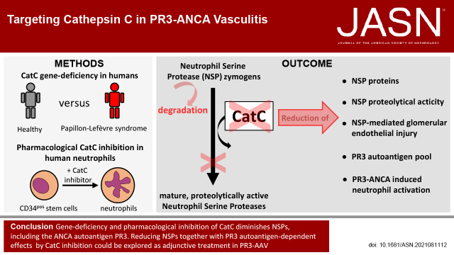
Background: The ANCA autoantigens proteinase 3 (PR3) and myeloperoxidase (MPO) are exclusively expressed by neutrophils and monocytes. ANCA-mediated activation of these cells is the key driver of the vascular injury process in ANCA-associated vasculitis (AAV), and neutrophil serine proteases (NSPs) are disease mediators. Cathepsin C (CatC) from zymogens activates the proteolytic function of NSPs, including PR3. Lack of NSP zymogen activation results in neutrophils with strongly reduced NSP proteins.

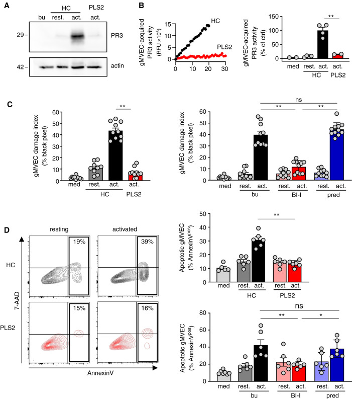
Methods: To explore AAV-relevant consequences of blocking NSP zymogen activation by CatC, we used myeloid cells from patients with Papillon-Lefèvre syndrome, a genetic deficiency of CatC, to assess NSPs and NSP-mediated endothelial cell injury. We also examined pharmacologic CatC inhibition in neutrophil-differentiated human hematopoietic stem cells, primary human umbilical vein cells, and primary glomerular microvascular endothelial cells.

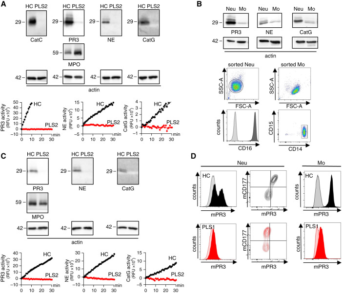
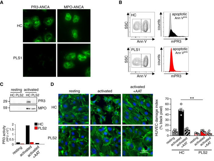
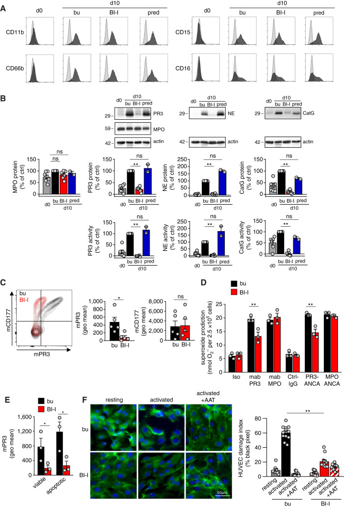
Results: Patients with Papillon-Lefèvre syndrome showed strongly reduced NSPs in neutrophils and monocytes. Neutrophils from these patients produced a negative PR3-ANCA test, presented less PR3 on the surface of viable and apoptotic cells, and caused significantly less damage in human umbilical vein cells. These findings were recapitulated in human stem cells, in which a highly specific CatC inhibitor, but not prednisolone, reduced NSPs without affecting neutrophil differentiation, reduced membrane PR3, and diminished neutrophil activation upon PR3-ANCA but not MPO-ANCA stimulation. Compared with healthy controls, neutrophils from patients with Papillon-Lefèvre syndrome transferred less proteolytically active NSPs to glomerular microvascular endothelial cells, the cell type targeted in ANCA-induced necrotizing crescentic glomerulonephritis. Finally, both genetic CatC deficiency and pharmacologic inhibition, but not prednisolone, reduced neutrophil-induced glomerular microvascular endothelial cell damage.

Conclusions: These findings may offer encouragement for clinical studies of adjunctive CatC inhibitor in patients with PR3-AAV.

Keywords: ANCA; cathepsin C; endothelial cells; immunology; vasculitis.


Figures

None
Graphical abstract
Figure 1.
Figure 1.
NSPs are strongly decreased in and on the surface of neutrophils and monocytes from patients with CatC gene-deficient PLS. (A) CatC, NE, PR3, and CatG in isolated blood neutrophils from an HC and a patient with PLS (PLS) by immunoblotting and by protease-specific FRET assays. MPO served as control and 42 kDa actin indicates equal sample loading. (B) NSP proteins in highly pure sorted HC monocytes (Mo) and neutrophils (Neu) by immunoblotting. Flow cytometry documents the purity of the sorted neutrophils and monocytes. (C) NSP protein expression and proteolytic activity in CD14hi sorted HC and PLS monocytes. (D) Flow cytometry of neutrophils stained for mPR3 and CD177. mPR3 expression on CD11bpos/CD14hi/CD15neg blood monocytes from an HC and a patient with PLS by flow cytometry.
Figure 2.
Figure 2.
PLS neutrophils produce a negative clinical PR3-ANCA immunofluorescence test, expose less PR3 on apoptotic neutrophils, and cause less NSP-mediated endothelial injury. (A) Indirect immunofluorescence using HC and PLS neutrophils and sera from patients on PR3 and MPO, respectively. (B) A percentage of overnight cultured neutrophils showed constitutive apoptosis by Annexin V staining. mPR3 was analyzed after gating on apoptotic (Annpos) cells. (C) cf-SN from resting and activated (2.5 µM ionophore A23187) HC and PLS neutrophils were assessed for PR3 protein by immunoblotting and proteolytic activity by FRET assay. (D) Confluent HUVEC monolayers were incubated with cf-SN from resting and activated HC and PLS neutrophils, respectively. When indicated cf-SN from activated neutrophils were treated with α1-antitrypsin (AAT) before EC incubation. EC damage was visualized by actin staining with phalloidin-FITC (green) and nuclear DAPI staining (blue), and quantified by determining the black pixel areas using a Leica microscope (×40) and ImageJ 1.48v software. ** P<0.01.
Figure 3.
Figure 3.
Pharmacologic CatC inhibition strongly reduces NSPs in neutrophils differentiated from HSC and subsequently reduces neutrophil activation by PR3-ANCA and EC injury. Neutrophils were differentiated from human CD34pos HSC for 10 days in the presence of buffer control (bu), BI-I, and prednisolone (pred) as indicated. (A) Neutrophil differentiation was assessed using the indicated surface markers. (B) NSP proteins were assessed by immunoblotting and the optical densities (OD) of the NSP bands were quantified. Proteolytic activity was measured by FRET assay. MPO served as a control. (C) Neutrophils differentiated for 10 days were double stained for mPR3 and CD177 and analyzed by flow cytometry. A typical experiment together with the mPR3 and CD177 mean fluorescence intensity are depicted. (D) Superoxide release by neutrophils differentiated for 10 days was assessed using mAbs to PR3 and MPO, human PR3- and MPO-ANCA IgG, and appropriate controls as indicated. (E) mPR3 on viable (AnnVneg) and apoptotic (Annpos) neutrophils differentiated for 10 days was assessed by flow cytometry. (F) The effect of cf-SN from resting and activated neutrophils differentiated for 10 days on EC injury was assessed by phalloidin-FITC (green) and nuclear DAPI staining (blue) and microscopy. *P<0.05 and **P<0.01.
Figure 4.
Figure 4.
Healthy control but not PLS neutrophils transfer proteolytically active PR3 to glomerular microvascular EC and both CatC gene deficiency and pharmacological CatC inhibition result in less neutrophil-induced glomerular microvascular EC injury. Confluent gMVEC monolayers were incubated with cf-SN from resting and activated HC or activated PLS neutrophils as indicated. After 1 hour, PR3 acquisition by glomerular microvascular EC was assessed by (A) immunoblotting using a PR3-specific mAb, and (B) FRET assay measuring proteolytic PR3 activity. Two independent experiments each with two HC and PLS2 neutrophils were performed. For assessing cell injury, glomerular microvascular EC monolayers were incubated with cf-SN from HC or PLS neutrophils, or from neutrophils that were differentiated from HSC in the presence of buffer, BI-I, or prednisolone as indicated. Each independent experiment includes neutrophils from two HC and PLS2, or two different HSC donors. Glomerular microvascular EC injury was determined (C) by phalloidin staining with the analysis of black pixel areas as in Figure 2D. The corresponding statistics is given (n=2), and (D) by Annexin V/7–AAD staining and flow cytometry (n=3). Contour plots of a typical experiment with cf-SN from resting and activated HC and PLS neutrophils are depicted together with the corresponding statistics of all experiments. *P<0.05 and **P<0.01.

Comment in

References

    1. van der Woude FJ, Rasmussen N, Lobatto S, Wiik A, Permin H, van Es LA, et al. : Autoantibodies against neutrophils and monocytes: tool for diagnosis and marker of disease activity in Wegener’s granulomatosis. Lancet 1: 425–429, 1985 - PubMed
    1. Falk RJ, Jennette JC: Anti-neutrophil cytoplasmic autoantibodies with specificity for myeloperoxidase in patients with systemic vasculitis and idiopathic necrotizing and crescentic glomerulonephritis. N Engl J Med 318: 1651–1657, 1988 - PubMed
    1. Lüdemann J, Utecht B, Gross WL: Anti-neutrophil cytoplasm antibodies in Wegener’s granulomatosis recognize an elastinolytic enzyme. J Exp Med 171: 357–362, 1990 - PMC - PubMed
    1. Goldschmeding R, van der Schoot CE, ten Bokkel Huinink D, Hack CE, van den Ende ME, Kallenberg CG, et al. : Wegener’s granulomatosis autoantibodies identify a novel diisopropylfluorophosphate-binding protein in the lysosomes of normal human neutrophils. J Clin Invest 84: 1577–1587, 1989 - PMC - PubMed
    1. Gupta SK, Niles JL, McCluskey RT, Arnaout MA: Identity of Wegener’s autoantigen (p29) with proteinase 3 and myeloblastin. Blood 76: 2162, 1990 - PubMed

Publication types

MeSH terms