. 2022 Mar 15;12(1):4462.
doi: 10.1038/s41598-022-08519-z.

The possible role of hypoxia in the affected tissue of relapsed clubfoot

Affiliations

The possible role of hypoxia in the affected tissue of relapsed clubfoot

Tomas Novotny et al. Sci Rep. .

Abstract

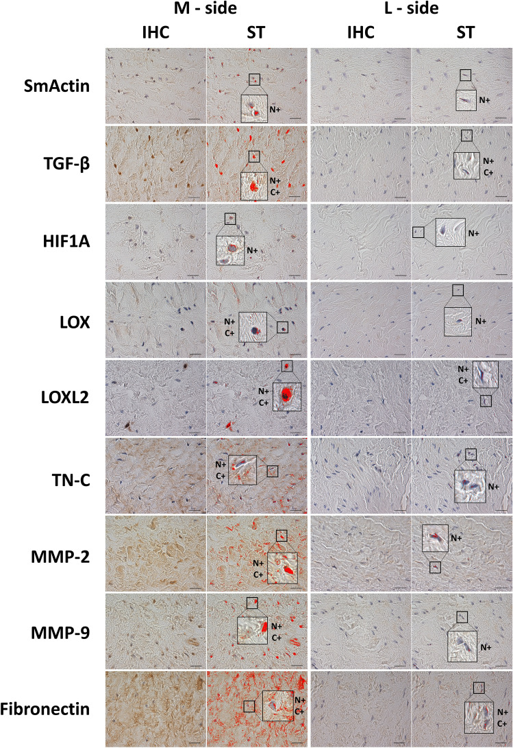
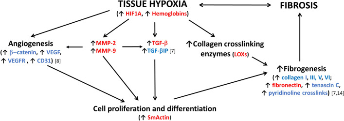
Our aim was to study the expression of hypoxia-related proteins as a possible regulatory pathway in the contracted side tissue of relapsed clubfoot. We compared the expression of hypoxia-related proteins in the tissue of the contracted (medial) side of relapsed clubfoot, and in the tissue of the non-contracted (lateral) side of relapsed clubfoot. Tissue samples from ten patients were analyzed by immunohistochemistry and image analysis, Real-time PCR and Mass Spectrometry to evaluate the differences in protein composition and gene expression. We found a significant increase in the levels of smooth muscle actin, transforming growth factor-beta, hypoxia-inducible factor 1 alpha, lysyl oxidase, lysyl oxidase-like 2, tenascin C, matrix metalloproteinase-2, matrix metalloproteinase-9, fibronectin, collagen types III and VI, hemoglobin subunit alpha and hemoglobin subunit beta, and an overexpression of ACTA2, FN1, TGFB1, HIF1A and MMP2 genes in the contracted medial side tissue of clubfoot. In the affected tissue, we have identified an increase in the level of hypoxia-related proteins, together with an overexpression of corresponding genes. Our results suggest that the hypoxia-associated pathway is potentially a factor contributing to the etiology of clubfoot relapses, as it stimulates both angioproliferation and fibroproliferation, which are considered to be key factors in the progression and development of relapses.


Conflict of interest statement

The authors declare no competing interests.

Figures

Figure 1
Figure 1
Demonstrative images of immunohistochemical staining and signal detection after the signal thresholding. Used antibodies: Anti-SmActin, Anti-TGF-β, Anti-HIF1A, Anti-LOX, Anti-LOXL2, Anti-TN-C, Anti-MMP-2, Anti-MMP-9, Anti-Fibronectin. The red areas in the ST columns represent the pixels analyzed after the thresholds were set. After applying the threshold settings, an image analyzer was used to measure the percentage of the area with a positive signal from the total area. IHC Immunohistochemistry, ST Signal Threshold, N+ Nuclear positivity, C+ Cytosolic positivity. Scale bar = 20 μm.
Figure 2
Figure 2
Immunohistochemical antibody detection. The percentage of (a) SmActin, TGF-β, HIF1A, LOX, LOXL2 and TN-C positive area and (b) the percentage of MMP-2, MMP-9 and Fibronectin positive area after immunohistochemical (IHC) detection. Data are presented as mean ± SD (n = 10). Values connected by lines differ significantly from each other (*p < 0.05, **p < 0.01, ***p < 0.005, ****p < 0.0005).
Figure 3
Figure 3
The fold change in the relative mRNA expression of selected genes (ACTA2, TGFB1, HIF1A, MMP2, FN1) in the M-side versus the L-side of the relapsed clubfoot tissue. A significant increase in expression was detected in the contracted medial side, in comparison with the non-contracted lateral side. The gene expression is normalized to the reference gene B2M and the mean expression value of the particular gene in the corresponding L-side tissue sample. Data are presented on a log scale as mean ± SD. The significance level is indicated as follows: *p < 0.05, **p < 0.01, ***p < 0.005.
Figure 4
Figure 4
Diagram of protein interactions involved in fibrosis formation in (relapsed) clubfoot. Marked proteins were found significantly upregulated (red—in this study; blue—in this study and/or in our other studies,,. SmActin alpha smooth muscle actin, HIF1A hypoxia-inducible factor 1 alpha, MMP-2, MMP-9 matrix metalloproteinase-2 and 9, LOXs lysyl oxidases, TGF-β transforming growth factor-beta, TGF-βIP transforming growth factor-beta-induced protein, VEGF vascular endothelial growth factor, VEGFR vascular endothelial growth factor receptor.

References

    1. Pavone V, et al. The etiology of idiopathic congenital talipes equinovarus: a systematic review. J. Orthop. Surg. Res. 2018;13:206. doi: 10.1186/s13018-018-0913-z. - DOI - PMC - PubMed
    1. Ansar A, et al. Systematic review and meta-analysis of global birth prevalence of clubfoot: a study protocol. BMJ Open. 2018;8:e019246. doi: 10.1136/bmjopen-2017-019246. - DOI - PMC - PubMed
    1. Dobbs MB, Gurnett CA. Update on Clubfoot: Etiology and treatment. Clin. Orthop. Relat. Res. 2009;467:1146–1153. doi: 10.1007/s11999-009-0734-9. - DOI - PMC - PubMed
    1. Hosseinzadeh P, Kiebzak G, Dolan L, Zionts L, Morcuende J. Management of clubfoot relapses with the ponseti method: Results of a survey of the POSNA members. J. Pediatr. Orthop. 2019;39:38–41. doi: 10.1097/BPO.0000000000000953. - DOI - PubMed
    1. Dobbs MB, et al. Factors predictive of outcome after use of the Ponseti method for the treatment of idiopathic clubfeet. J. Bone Joint. Surg. Am. 2004;86:22–27. doi: 10.2106/00004623-200401000-00005. - DOI - PubMed

Publication types

Substances