Disease correlates of rim lesions on quantitative susceptibility mapping in multiple sclerosis
- PMID: 35292734
- PMCID: PMC8924224
- DOI: 10.1038/s41598-022-08477-6
Disease correlates of rim lesions on quantitative susceptibility mapping in multiple sclerosis
Abstract
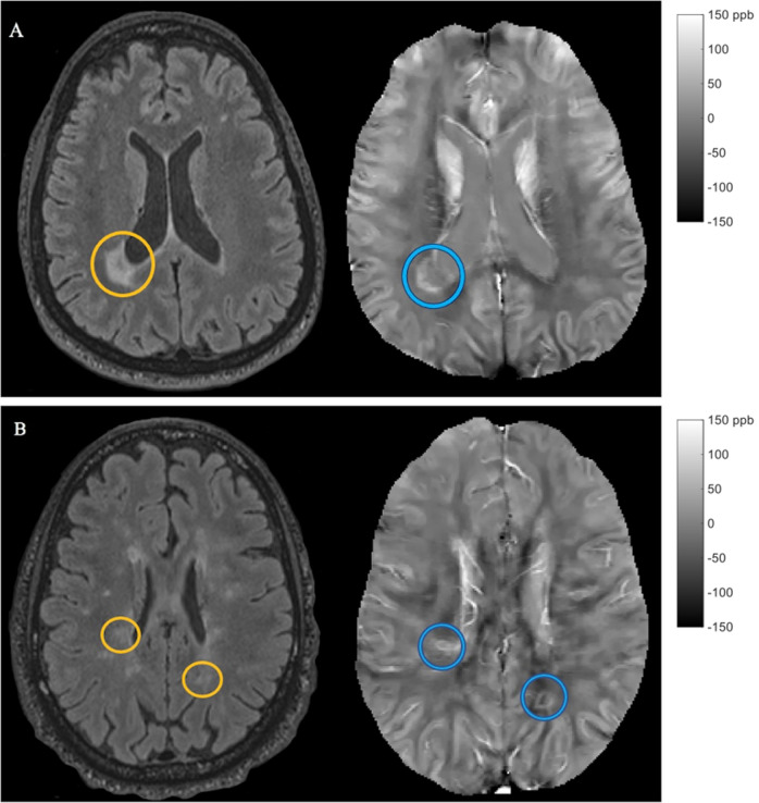
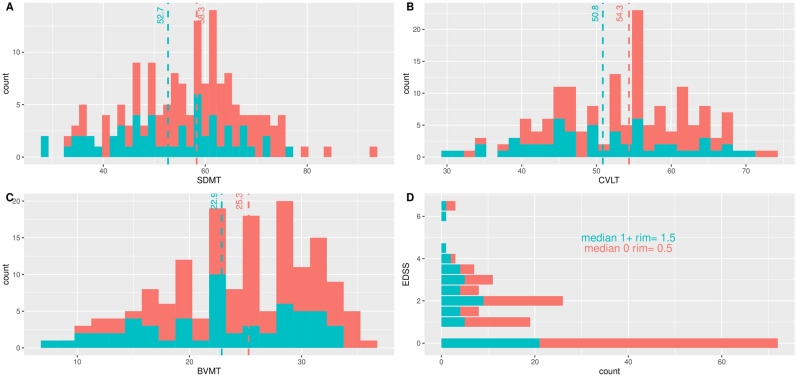
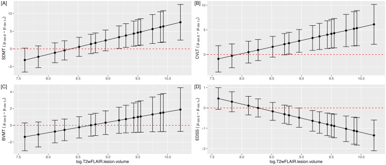
Quantitative susceptibility mapping (QSM), an imaging technique sensitive to brain iron, has been used to detect paramagnetic rims of iron-laden active microglia and macrophages in a subset of multiple sclerosis (MS) lesions, known as rim+ lesions, that are consistent with chronic active lesions. Because of the potential impact of rim+ lesions on disease progression and tissue damage, investigating their influence on disability and neurodegeneration is critical to establish the impact of these lesions on the disease course. This study aimed to explore the relationship between chronic active rim+ lesions, identified as having a hyperintense rim on QSM, and both clinical disability and imaging measures of neurodegeneration in patients with MS. The patient cohort was composed of 159 relapsing-remitting multiple sclerosis patients. The Expanded Disability Status Scale (EDSS) and Brief International Cognitive Assessment for Multiple Sclerosis, which includes both the Symbol Digit Modalities Test and California Verbal Learning Test-II, were used to assess clinical disability. Cortical thickness and thalamic volume were evaluated as imaging measures of neurodegeneration. A total of 4469 MS lesions were identified, of which 171 QSM rim+ (3.8%) lesions were identified among 57 patients (35.8%). In a multivariate regression model, as the overall total lesion burden increased, patients with at least one rim+ lesion on QSM performed worse on both physical disability and cognitive assessments, specifically the Symbol Digit Modalities Test (p = 0.010), California Verbal Learning Test-II (p = 0.030), and EDSS (p = 0.001). In a separate univariate regression model, controlling for age (p < 0.001) and having at least one rim+ lesion was related to more cortical thinning (p = 0.03) in younger patients (< 45 years). Lower thalamic volume was associated with older patients (p = 0.038) and larger total lesion burden (p < 0.001); however, the association did not remain significant with rim+ lesions (p = 0.10). Our findings demonstrate a novel observation that chronic active lesions, as identified on QSM, modify the impact of lesion burden on clinical disability in MS patients. These results support further exploration of rim+ lesions for therapeutic targeting in MS to reduce disability and subsequent neurodegeneration.
© 2022. The Author(s).
Conflict of interest statement
Dr. Wang owns equity of Medimagemetric LLC. Dr. Gauthier reported receiving grants from Genentech, Sanofi-Genzyme, and Mallinckrodt outside the submitted work. No other disclosures were reported.
Figures