Review
. 2022 Mar 15;24(2):43.
doi: 10.1208/s12248-022-00692-3.

Nanotechnology-Driven Cell-Based Therapies in Regenerative Medicine

Affiliations
Review

Nanotechnology-Driven Cell-Based Therapies in Regenerative Medicine

D Alzate-Correa et al. AAPS J. .

Abstract

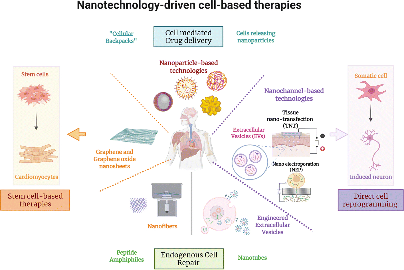
The administration of cells as therapeutic agents has emerged as a novel approach to complement the use of small molecule drugs and other biologics for the treatment of numerous conditions. Although the use of cells for structural and/or functional tissue repair and regeneration provides new avenues to address increasingly complex disease processes, it also faces numerous challenges related to efficacy, safety, and translational potential. Recent advances in nanotechnology-driven cell therapies have the potential to overcome many of these issues through precise modulation of cellular behavior. Here, we describe several approaches that illustrate the use of different nanotechnologies for the optimization of cell therapies and discuss some of the obstacles that need to be overcome to allow for the widespread implementation of nanotechnology-based cell therapies in regenerative medicine.

Keywords: direct cell reprogramming; nanotechnology; nanotransfection; regenerative medicine; stem cell therapy.


Conflict of interest statement

Conflict of Interest Statement:

The authors have no conflicts of interest to declare.

Figures

Figure 1.
Figure 1.
Nanotechnology-based strategies to improve safety and efficacy of stem cell- and direct reprogramming-based cell therapies.
Figure 2.
Figure 2.. Approaches employing graphene oxide nanosheets and nanofibers to regulate stem cell behavior.
(A) Schematic representation of the experimental design to evaluate the response of hemangioblasts to gelatin (GE) or graphene oxide (GO). Heaemanglioblast cultures on GE/GO showing higher (B and D) GFP signal and (C and E) production of CD41+ / GFP− cells on GO-coated coverslips compared to GE (Adapted from Garcia-Alegria et al., 2016. Ref 14). Expression of sarcomeric alpha-actinin (α-SA), a cardiomyocyte marker, in human iPSCs (hiPSCs) differentiated using gelatin-PCL nanofiber scaffolds in a (F) 2D and (G) 3D culture system. (H) Gene expression of cardiac progenitor and cardiomyocyte markers SIRPA/ISL1, MHC6/TNNT2, respectively during differentiation of hiPSCs into cardiomyocytes in 2D and 3D cultures (Adapted from Sridharan et al., 2021. Ref 27).
Figure 3.
Figure 3.. Methodologies using nanoparticles to modulate stem cell differentiation.
(A) Properties of nanoparticles (NPs) loaded with microRNAs. (B) Experimental setup where mice were subjected to two stereotaxic injections, one in the right striatum to deliver 6-Hydroxydopamine (6-OHDA) to induce a PD phenotype, and another in the right lateral ventricle to deliver miR-124 NPs or saline solution. (C-D) Confocal images of BrdU (proliferation marker, green), Hoechst (nuclear marker, blue), and DCX (mature neuronal marker, red) staining showing an increase in the number of mature neurons (NeuN+/BrdU+ cells) observed in the striatum of mice treated with 6-OHDA and miR-124 NPs compared to healthy controls. (E) Apomorphine-rotation test (behavioral analysis) illustrates a decrease in motor deficits (net contralateral rotations) in mice treated with miR-124 NP (Adapted from Saraiva et al., 2016. Ref 37). (F) Composition of the traceable NPs PHEMA-RA-PCB-CPP/SPIONs/siSOX9 (condensed as ABC/SPIONs/siSOX9 NPs: S8). (G) Immunostaining analysis with MAP-2 (neuronal marker, green), GFAP (glial cell marker, red), and DAPI (nuclear marker, blue), showing higher MAP-2 expression (conversion into neurons) when treated with S8 compared to control. (H) Morris water maze experiments were performed to assess the effect of NP treatment on spatial learning and memory improvement, showing that NSCs treated with S8 NPs could potentially improve cognition and memory (Adapted from Zhang et al., 2016. Ref 40).
Figure 4.
Figure 4.. Approaches used for endogenous cell repair
(A) Representation of the chemical structure of the PA intended to mirror VEGF’s activity. (B) Nanofiber structure formed by the VEGF can be seen by cryogenic Transmission electron microscopy (TEM) (C), as well as the interconnected nanofiber gel network, captured by Scanning electron microscopy (SEM). To evaluate the ability of VEGF-mimetic PA as a therapy for ischemic disorders using a murine hind-limb ischemia model, limb salvage and motor function was evaluated, showing (D) an improvement in tissue salvage score (i.e., less necrosis) and (E) a significant effect on active limb motor function in the treatment group compared to the control groups. (F) Functional tests show enhanced walking time preceding failure, and (G-H) Laser Doppler perfusion imaging shows enhanced tissue perfusion ratio in the ischemic hind limb for 28 days following treatment (Adapted from Webber et al., 2011. Ref 44). High concentration of methyl acrylic anhydride-gelatin (GelMA)-Ppy nanoparticles were used to fabricate engineered cardiac patches (ECP). Characterization and analysis of the nanoparticles, showing (I) uniform spherical morphology and size via TEM, and (J) molecular structure via X-ray diffraction (XRD). (K) Live/death staining shows great biocompatibility of the nanoparticles for 7 days without affecting cell growth. (L) Cardiac sections were stained with Masson’s staining for fibrous tissue (blue) and myocardium (red), showing enhanced cardiac function and revascularization for patch-implanted groups, which is also evident in the analysis of (M) the infarct size and (N) the infarct wall thickness (Adapted from He et al., 2018. Ref 51).
Figure 5.
Figure 5.. Methodologies employed for cell-mediated drug delivery.
(A) Schematic of the structure of the cellular backpack, loaded with catalase, showing the composition and assembly of different regions from the “release region” to the cell attachment region. (B) Confocal microscopy images of the whole brain after systemic delivery of backpack-carrying macrophages show fluorescently labeled macrophages (green) and backpacks (red). (C) At 40X magnification, co-localization of green and red can be observed, suggesting that the cells facilitated the transport of backpacks to the brain (Adapted from Klyachko et al., 2017. Ref 53). (D) Systemic delivery of bone marrow-derived macrophages (BMM) loaded with a catalase nanozyme in a murine model of brain inflammation shows an increased blood concentration of nanozyme for more than 170 hours after injection. (E) Increased accumulation of catalase is found in all tissues (brain, spleen, liver, and Kidney) when using nanozyme-loaded BMM. Higher accumulation of the nanozyme is found in the spleen and lower accumulation in the brain. (F) Biodistribution of BMM loaded with fluorescently labeled nanozyme, showing targeted drug delivery from peripheral organs to the brain with inflammation for over 16–20 days (top panel) compared to healthy animals (bottom panel) (Adapted from Zhao et al., 2011. Ref 54).
Figure 6.
Figure 6.. Approaches implementing nanoparticles for the direct reprogramming of somatic cells.
(A) Induced hepatocyte-like cells (iHeps) converted from mouse fibroblasts using MSN/PEI/Transcription factor nanocomplexes. (B) Successful conversion into iHeps by quantitative expression, showing gradual downregulation of Col1a1, Desmin and Fsp1 (fibroblasts genes) and upregulation of hepatocyte genes (rest of the genes) in the treatment group compared to other groups (Adapted from Wang et al., 2020. Ref 61). (C) Schematic illustration of direct injection of PEI/gold nanoparticles loaded with GMT genes (AuNP/GMT/PEI) into the heart of a mouse. (D) Cell cytometry analysis showing efficient reprogramming from mouse embryonic fibroblasts into induced cardiomyocytes after using AuNP/GMT/PEI nanocomplexes by the expression of αMHC. (E) Upregulation of cardiomyocyte genes in the treatment group compared to control via qRT-PCR. (F) immunostaining of injured heart displaying a significant increase in the number of cardiac Troponin T + (cTNT) and α-MHC+ cells relative to controls (Adapted from Chang et al., 2019. Ref 62).
Figure 7.
Figure 7.. Nanochannel-based technology to modulate the direct reprogramming of fibroblasts into induced endothelial cells as therapeutic agents for stroke.
(A) Schematic diagram illustrating the nanotransfection with EFF, release of pro-vasculogenic/angiogenic EVs (e.g., exosomes), and reprogramming of fibroblasts into induced endothelial cells (iECs) that subsequently mediate the formation of induced vasculature (iVas). (B) Upregulation of genes in nanotransfected cells and (C) loaded in released exosomes. (D) In vitro tube formation assay in the EFF group. (E) Schematic diagram of middle cerebral artery occlusion, intracranial injection, and MRIs. (F, G) T2-weighted MR images post-stroke show that intracranial injection of EFF-nanotransfected cells led to significantly improved infarct resolution compared to control in mice that exceeded 17% weight loss. EFF-nanotransfected cells injected in mice show (H) superior neuronal cellularity (NeuN) and (I) reduced astroglial scar formation (GFAP) (Adapted from Lemmerman et al., 2021. Ref 68).
Figure 8.
Figure 8.. Methodologies employing nanochannel-based technologies to regulate the direct reprogramming of fibroblasts into neurons in vivo and promote nerve tissue repair.
(A) Schematic representation of the TNT procedure on the skin, where an electric field is applied through the electrodes to create nanopores in the membranes of exposed cells and drive cargo into the skin-cells via electrophoresis. (B) The outermost cell layer is in direct contact with the Nanochannels. (C) Simulations show focused (solid) compared to widespread (dashed) poration in TNT vs. Bulk electroporation (BEP). (D) Mouse skin showing successful gene delivery and expression via confocal imaging. (E) Epidermis and dermis analyses showing gene expression using Laser capture microdissection (LCM) and qRT–PCR. Immunostaining results display increased (F) TUJ1 and (G) neurofilament (NF) expression after TNT-based delivery of ABM. (H) TNT with EFF on the skin of mice led to (I-J) increased iVAS (Pecam-1, vWF) at day 7 (Adapted from Gallego-Perez et al., 2017. Ref 66). (K) Delivery of EFF using TNT in a crushed nerve tissue model, which leads to (L-M) increased vascularity, as well as (N) accelerated recovery of nerve function, which was assessed using compound muscle action potential (CMAP) measurements (Adapted from Moore et al., 2020. Ref 69).

References

    1. Wang M-L. The Modern Pharmaceutical Industry: History, Current Position and Challenges. Global Health Partnerships: The Pharmaceutical Industry and BRICA. London: Palgrave Macmillan UK; 2009. p. 33–80.
    1. Mason C, Brindley DA, Culme-Seymour EJ, Davie NL. Cell therapy industry: billion dollar global business with unlimited potential. Regen Med. 2011;6(3):265–72. - PubMed
    1. Fischbach MA, Bluestone JA, Lim WA. Cell-based therapeutics: the next pillar of medicine. Sci Transl Med. 2013;5(179):179ps7. - PMC - PubMed
    1. Au P, Hursh DA, Lim A, Moos MC Jr., Oh SS, Schneider BS, et al. FDA oversight of cell therapy clinical trials. Sci Transl Med. 2012;4(149):149fs31. - PubMed
    1. Towards advanced cell therapies. Nature Biomedical Engineering. 2018;2(6):339–40. - PubMed

Publication types

LinkOut - more resources