

Preserving mitochondrial function by inhibiting GRP75 ameliorates neuron injury under ischemic stroke
- PMID: 35293600
- PMCID: PMC8941507
- DOI: 10.3892/mmr.2022.12681
Preserving mitochondrial function by inhibiting GRP75 ameliorates neuron injury under ischemic stroke
Abstract
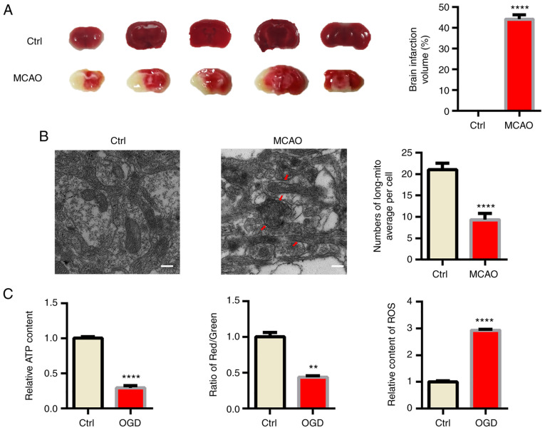
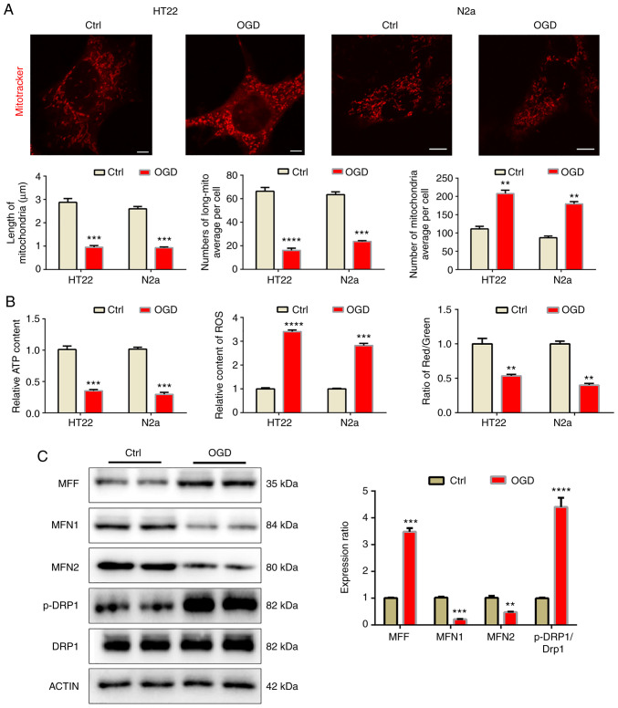
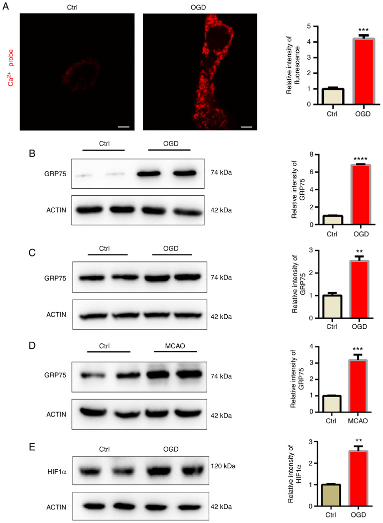
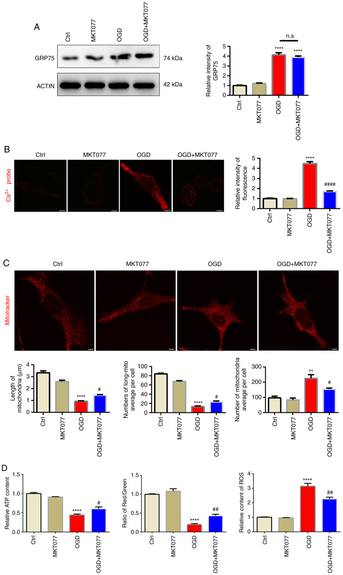
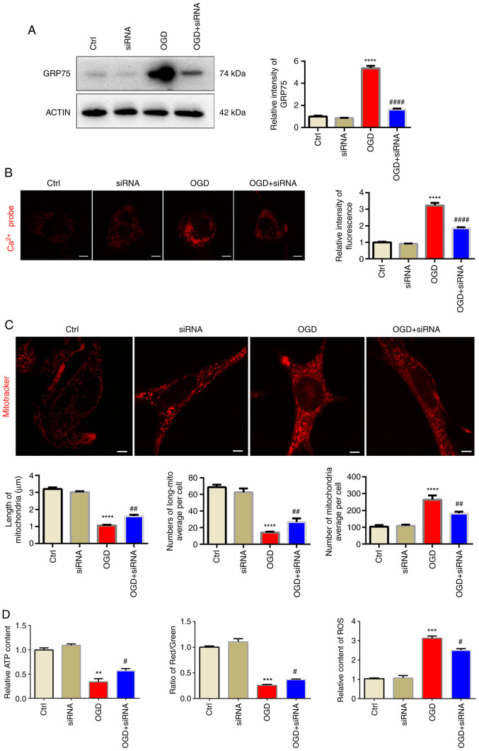
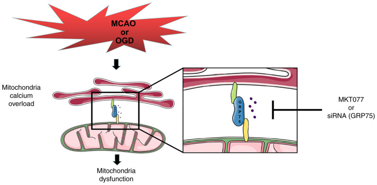
Ischemic stroke is a life‑threatening disease, which is closely related to neuron damage during ischemia. Mitochondrial dysfunction is essentially involved in the pathophysiological process of ischemic stroke. Mitochondrial calcium overload contributes to the development of mitochondrial dysfunction. However, the underlying mechanisms of mitochondrial calcium overload are far from being fully revealed. In the present study, middle cerebral artery obstruction (MCAO) was performed in vivo and oxygen and glucose deprivation (OGD) in vitro. The results indicated that both MCAO and OGD induced significant mitochondrial dysfunction in vivo and in vitro. The mitochondria became fragmented under hypoxia conditions, accompanied with upregulation of the heat shock protein 75 kDa glucose‑regulated protein (GRP75). Inhibition of GRP75 was able to effectively ameliorate mitochondrial calcium overload and preserve mitochondrial function, which may provide evidence for further translational studies of ischemic diseases.
Keywords: GRP75; calcium overload; mitochondria; mitochondria‑associated endoplasmic reticulum membrane.
Conflict of interest statement
The authors declare that they have no competing interests.
Figures
Similar articles
-
Potential targets for the treatment of MI: GRP75-mediated Ca2+ transfer in MAM.Eur J Pharmacol. 2024 May 15;971:176530. doi: 10.1016/j.ejphar.2024.176530. Epub 2024 Mar 23. Eur J Pharmacol. 2024. PMID: 38527700
-
Up-regulation of miR-122 protects against neuronal cell death in ischemic stroke through the heat shock protein 70-dependent NF-κB pathway by targeting FOXO3.Exp Cell Res. 2018 Aug 1;369(1):34-42. doi: 10.1016/j.yexcr.2018.04.027. Epub 2018 Apr 30. Exp Cell Res. 2018. PMID: 29715465
-
IP3R-Grp75-VDAC1-MCU calcium regulation axis antagonists protect podocytes from apoptosis and decrease proteinuria in an Adriamycin nephropathy rat model.BMC Nephrol. 2018 Jun 15;19(1):140. doi: 10.1186/s12882-018-0940-3. BMC Nephrol. 2018. PMID: 29907098 Free PMC article.
-
Role of Calcium Homeostasis in Ischemic Stroke: A Review.CNS Neurol Disord Drug Targets. 2022;21(1):52-61. doi: 10.2174/1871527320666210212141232. CNS Neurol Disord Drug Targets. 2022. PMID: 33583386 Review.
-
Role of mitochondrial metabolism in ischemic stroke and natural products intervention.Mol Biol Rep. 2025 Jun 7;52(1):568. doi: 10.1007/s11033-025-10667-0. Mol Biol Rep. 2025. PMID: 40481903 Review.
Cited by
-
Reduction of Mitochondrial Calcium Overload via MKT077-Induced Inhibition of Glucose-Regulated Protein 75 Alleviates Skeletal Muscle Pathology in Dystrophin-Deficient mdx Mice.Int J Mol Sci. 2024 Sep 13;25(18):9892. doi: 10.3390/ijms25189892. Int J Mol Sci. 2024. PMID: 39337383 Free PMC article.
-
Stroke: Molecular mechanisms and therapies: Update on recent developments.Neurochem Int. 2023 Jan;162:105458. doi: 10.1016/j.neuint.2022.105458. Epub 2022 Nov 30. Neurochem Int. 2023. PMID: 36460240 Free PMC article. Review.
-
Calcium bridges built by mitochondria-associated endoplasmic reticulum membranes: potential targets for neural repair in neurological diseases.Neural Regen Res. 2025 Dec 1;20(12):3349-3369. doi: 10.4103/NRR.NRR-D-24-00630. Epub 2024 Nov 13. Neural Regen Res. 2025. PMID: 39589178 Free PMC article.
-
CaM promotes cardiomyocyte mitophagy in myocardial ischemia-reperfusion injury involving in the regulation of the IP3R3-GRP75-VDAC1 complex.Sci Rep. 2025 Jul 1;15(1):22379. doi: 10.1038/s41598-025-07977-5. Sci Rep. 2025. PMID: 40595218 Free PMC article.
-
The Therapeutic Effects of Blueberry-Treated Stem Cell-Derived Extracellular Vesicles in Ischemic Stroke.Int J Mol Sci. 2024 Jun 8;25(12):6362. doi: 10.3390/ijms25126362. Int J Mol Sci. 2024. PMID: 38928069 Free PMC article.
References
MeSH terms
Substances
LinkOut - more resources
Full Text Sources
Medical
